|
Carman Meniscus Sign-large gastric ulcer seen on UGI convex in towards the lumen of the stomach, the rolled edges indicative of malignancy |
|
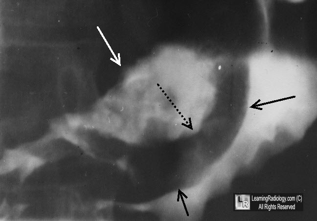
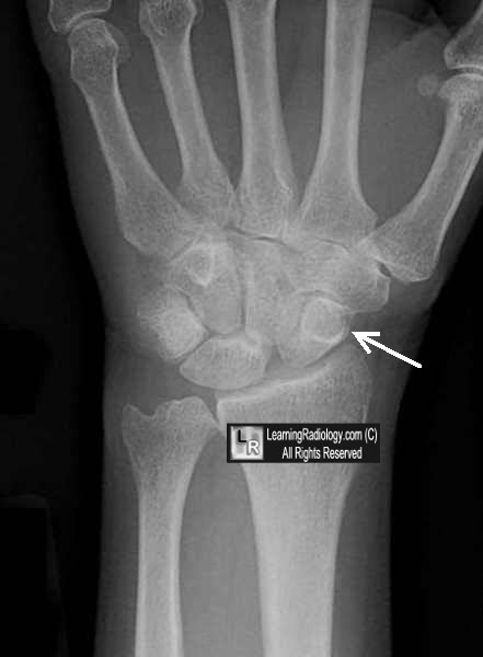
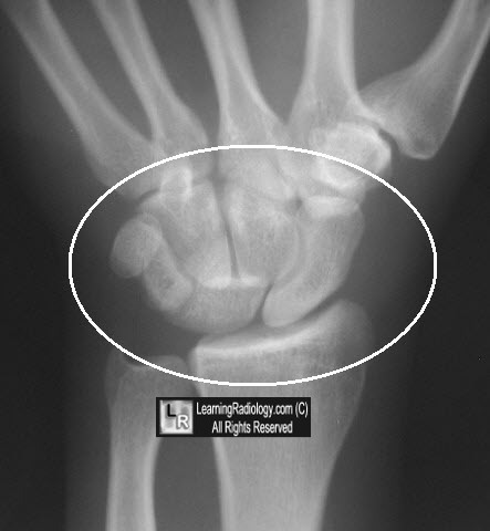
Cortical Ring Sign-circular shadow cast by rotated scaphoid in scapholunate subluxation secondary to scaphoid's abnormal orientation |
|
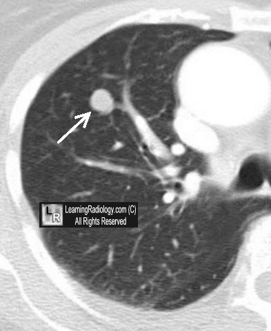
Coin Lesion-a solitary pulmonary nodule generally considered less than 3 cm in size, most often a granuloma or hamartoma |
|
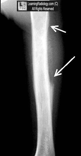
Codman's Triangle-triangular elevation of periosteum from an aggressive, usually malignant, bone tumor such as an osteosarcoma |
|
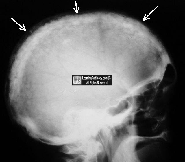
Cotton Wool Appearance-islands of poorly marginated sclerotic disease surrounded by less dense skull in Paget disease |
|
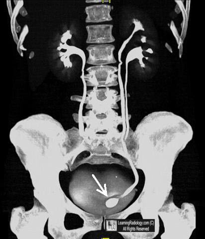
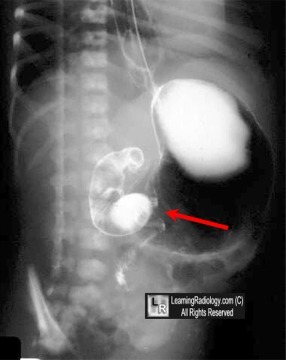
Cobra Head Abnormality-rounded dilatation of distal ureter, surrounded by thin lucent line, seen in patients with adult-type ureteroceles |
|
Cobblestone Appearance-GI-alternating normal and denuded mucosa from ulceration, esp in Crohn's disease of the colon |
|
C Sign-on lateral view of foot in tarsal coalition, a continuous arc from medial cortex of talus to inferior cortex of sustentaculum talus |
|
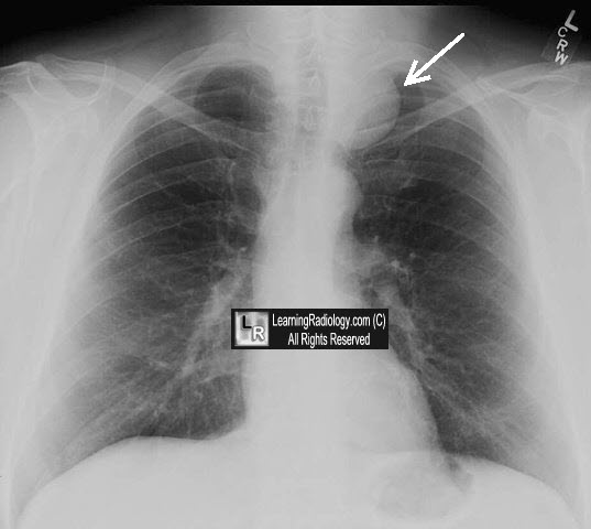
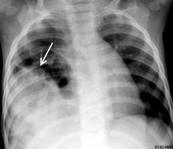
Cervicothoracic Sign-a mass extends above clavicles on frontal chest radiograph should be posterior in chest |
|
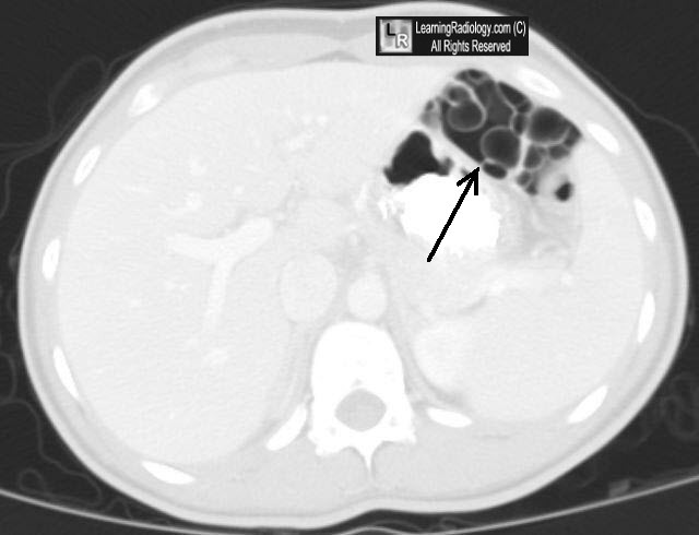
Cluster of Grapes Sign-multiple gas-containing cysts usually in segment of left colonic wall in pneumatosis cystoides intestinalis |
|
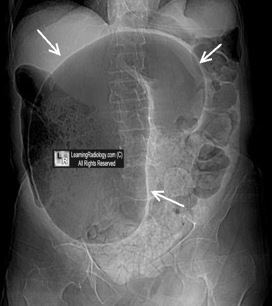
Coffee-bean Sign-dilated sigmoid colon in sigmoid volvulus thought to resemble a giant coffee-bean |
|
Colon Cutoff Sign-dilated transverse colon, usually to splenic flexure, associated with pancreatitis or ischemic colitis |
|
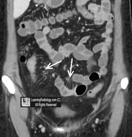
Comb Sign-contrast-enhanced tubular
opacities on the mesenteric side of the ileum aligned like the teeth of a comb, especially in Crohn disease |
|
Comet Tail Sign-lung-focal area of collapsed lung adjacent to pleural thickening with distortion of blood vessels in rounded atelectasis |
|
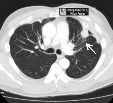
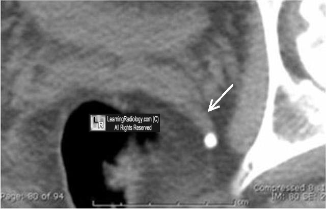
Comet Sign-appearance of a calcified phlebolith and its adjacent non-calcified vein; may aid in DDX from ureteral calculus |
|
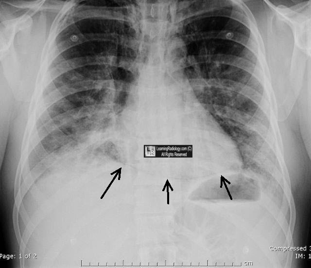
Continuous Diaphragm Sign-visualization of entire upper surface of diaphragm from pneumomediastinum |
|
Corduroy Sign-vertically oriented, thickened trabeculae seen in vertebral body hemangiomas |
|
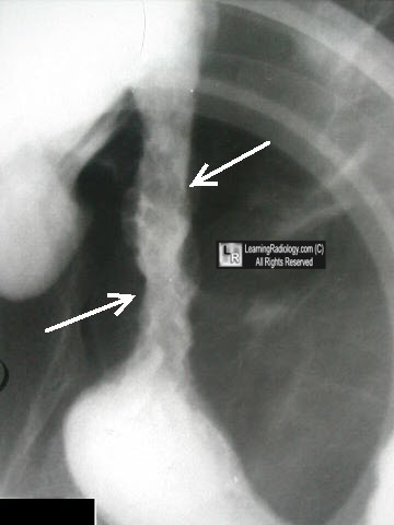
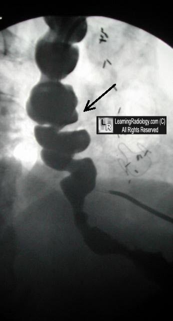
Corkscrew Esophagus-constricted, twisted lumen usually seen in diffuse esophageal spasm from abnormal, tertiary contractions |
|
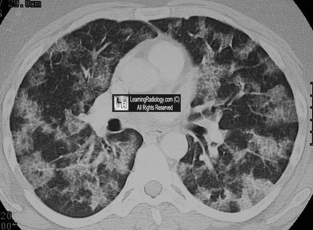
Crazy Paving Sign-fine reticular pattern superimposed on areas of ground-glass opacity on HRCT, first described with alveolar proteinosis |
|
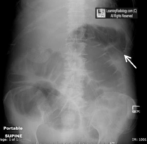
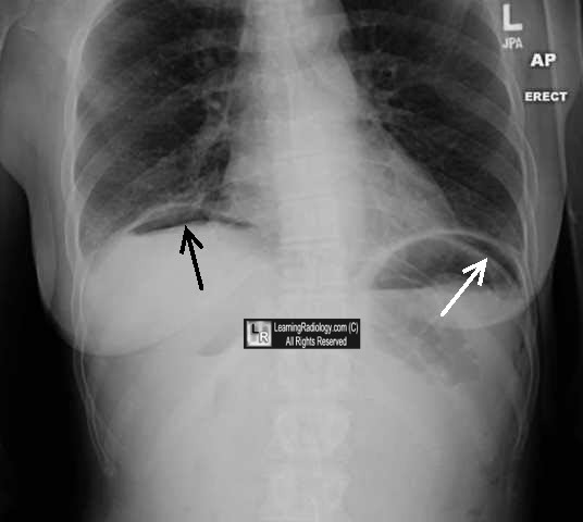
Crescent Sign-appearance of a sliver of air usually best seen beneath the right hemidiaphragm in pneumoperitoneum |
|
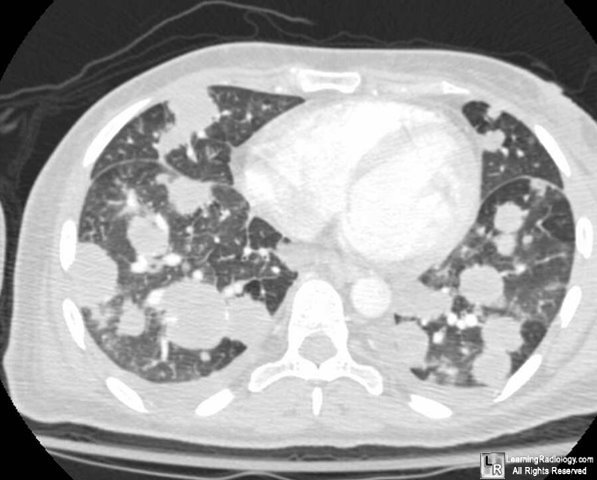
Cannonball Metastases-large, hematogenously spread metstatic lesions in the lungs of varying sizes most often from colon, breast, renal, thyroid primaries |
|
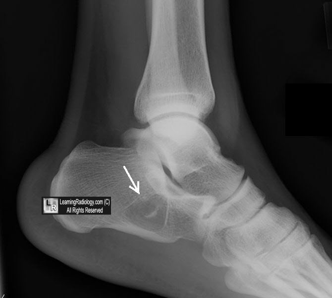
Cockade Sign-intraosseous calcaneal lipoma with central calcification resembling the badge generally worn upon a hat |
|
Collar Button Ulcer-GI-ulceration with undermining of submucosa producing distinctive shape, esp with ulcerative colitis |
|
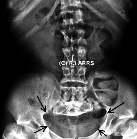
Champagne Glass Pelvis-squaring of the iliac wings in achondroplasia producing a champagne glass appearance of the pelvic inlet |
|
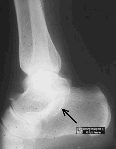
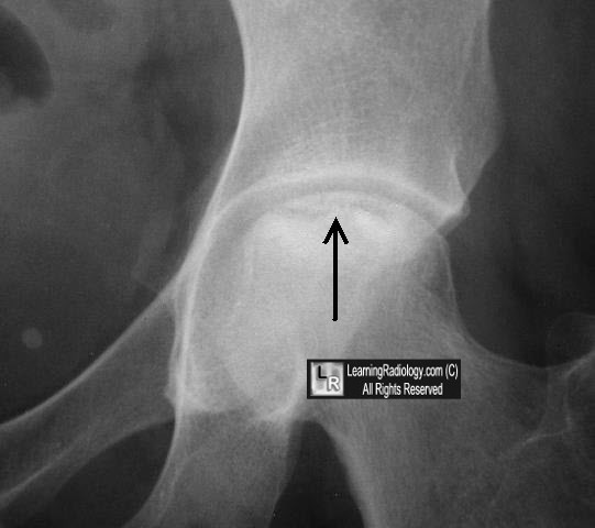
Crescent Sign (Hip)--subarticular radiolucency of femoral head seen best on frog lateral in avascular necrosis |
|
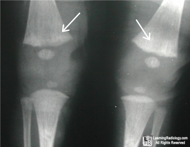
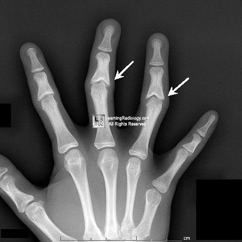
Celery Stalking-striated appearance of metaphyses in patients with rubella and osteopathia striata; also degenerated ACL on MRI |
|
Corkscrew Sign-spiral appearance of 4th part of duodenum on UGI in children with mid-gut volvulus |
|
Cottage Loaf Sign-constricted appearance of liver herniated through a right-sided diaphragmatic rupture looks like English cottage loaf |
|
Coned Epiphyses-are usually normal but may occur in sickle cell anemia, congenital diseases, infection, trauma, and radiation injury |
|
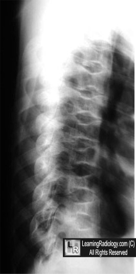
Codfish Vertebra-biconcave appearance of the vertebral bodies themselves in sickle cell disease from avascular necrosis resembling the vertebra of codfish |
|
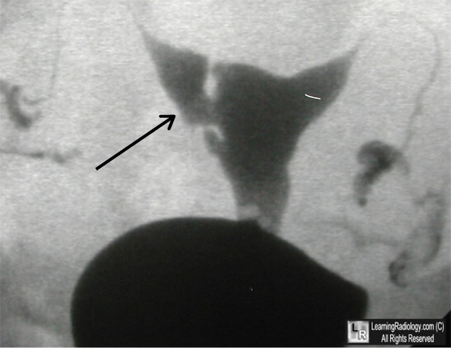
Crowded Carpal Sign-overlap of the distal and proximal carpal rows in perilunate dislocation |
|
Cupola Sign-arcuate lucency superimposed on lower thoracic spine on a supine abdomen representing free air under the central diaphragmatic tendon |
|
Cobblestone Pattern-hysterosalpingography-rounded filling defects from intraluminal adhesions |