|
|
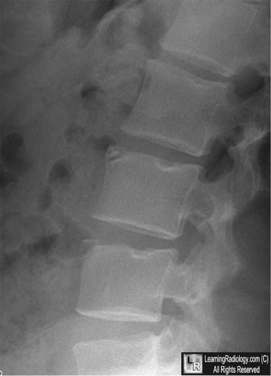
Limbus Vertebra
General Considerations
- Common finding
- Most commonly affects the anterosuperior corner of vertebral body in a single vertebral body
- May also affect the inferior margin less commonly and posterior margins least commonly
- Seen mostly in mid-lumbar spine
- Probably due to trauma
- Caused by herniation of a portion of the nucleus pulposus underneath the ring apophysis before its fusion to the body
- Ring apophysis stays separate from the main body
Clinical Findings
- Posterior limbus vertebra have been reported to cause nerve compression
- Anterior limbus vertebra are not believed to cause symptoms
Imaging Findings
- Triangular fragment of bone at anterosuperior corner of a vertebral body
- Sclerotic margins to fragment
- Sclerotic margin of adjacent vertebral body
Differential Diagnosis
- Fracture

- Typically do not have sclerotic margins like limbus vertebra
- Discitis
- Especially in children where a limbus vertebra may not have a sclerotic margin
- Schmorl's node
- More central defect also caused by herniation of nucleus pulposus
Treatment
Complications
|
|
Limbus vertebra |
Limbus vertebra |
|
|
Discitis |
Fracture |
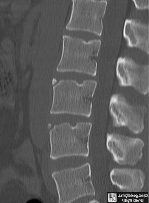
Limbus vertebra (and look-alikes). The limbus vertebra (top) shows corner fragments that
are well-corticated (white arrows) The same bodies contain Schmorl's nodes (yellow arrows). Discitis (lower left)
destroys two adjacent endplates (yellow and white arrows) and the intervening disk space. The edges are not sclerotic.
The fracture has non-sclerotic margins (white arrow) and extends through the vertebral body (blue arrow).
For more information, click on the link if you see this icon 
For these same photos without the annotations, click here and here
The Limbus Vertebra: An Anterior Disc Herniation Demonstrated by Discography. Bernard Ghelman And Robert H. Freiberger. Am J Roentgenol 127:854-855, 1976
|
|
|


