|
|
Planum Sphenoidale Meningioma
General Considerations
- As many as 10% of meningiomas
- Typically occur in women in 5th and 6th decades of life
- Occur at the anterior cranial base overlying the cribiform plate of the ethmoid bone, frontosphenoid suture and planum sphenoidale
- Meningiomas are extra-axial lesions that arise from arachnoid cells
- Patients with neurofibromatosis type 2 (NF-2) have a 50% chance of developing one or more meningiomas
Clinical Findings
- Often attain significant size before producing symptoms
- Changes in cognition and personality
- Headaches
- Visual disturbances, often more severe ion one eye but both are commonly involved
- Seizures
Imaging Findings
- CT
- CT is important for showing bony involvement which may be helpful in surgical planning
- On unenhanced studies. They are isodense to slightly hyperdense
- With contrast, they enhance intensely and homogeneously
- There may be extensive surrounding edema
- Adjacent brain is compressed but not invaded
- MRI
- They have variable signal intensity on T1 and T2-weighted images
- After gadolinium, they again enhance homogeneously and intensely
- More perilesional edema may be seen than on CT
- At their periphery, an enhancing tail to the dura may be seen
- Cystic meningiomas may exhibit intratumoral or peritumoral cysts
Differential Diagnosis
- Multiple meningiomas may resemble metastases
Treatment
- Surgical resection
- Radiation therapy
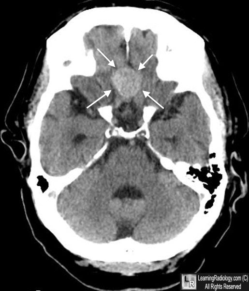
Planum Sphenoidale Meningioma. Unenhanced axial CT of the base of the skull shows a hyperdense midline mass (white arrows) arising from the planum sphenoidale region.
Meningiomas of the Tuberculum Sellae and Planum Sphenoidale: A Review of 83 Cases. JE Finn, LA Moun. Arch Ophthalmol. 1974;92(1):23-27
|
|
|
