1. Arthritis A practical radiological approach


Arthritis — narrowing the radiographicdiagnosis
Distribution of disease
Age and sex
Look for characteristic radiologicalfindings
Associated clinical illness?
Osteoarthritis — hallmarks
Primary defect is loss of cartilage
Leads to asymmetric joint space narrowing
Bone production:
Marginal osteophytes
Subchondral sclerosis
Subchondral cysts
Loose bodies (“joint mice”)
Arthritis — non-inflammatory
Osteoarthritis
Large joints
Small joints
Post-traumatic arthritis
Neuropathic arthritis
Arthritis — inflammatory
Rheumatoid arthritis
Gout
CPPD deposition disease
Septic arthritis
Seronegitive spondylarthropathies
Reiter’s
Psoriatic arthritis
Ankylosing spondylitis
Normal joints
Normal joints
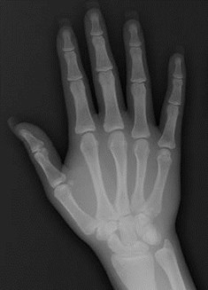
Primary Osteoarthritis
= Degenerative joint disease
Idiopathic disease of small joints,especially hand and wrist
Chronic degenerative disease oflarge weight bearing joints especiallyspine, hips and knees
Weight bearing joint spaces involvedfirst and worst
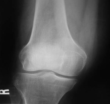
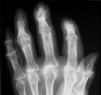
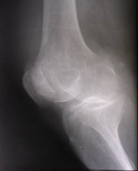
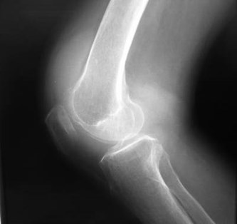
Osteoarthritis—hypertrophic spurring
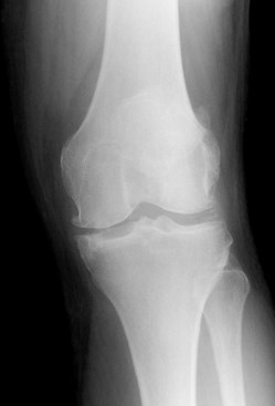
Osteoarthritis — progressive jointspace narrowing
Also progressive sclerosis andsubchondral cyst formation
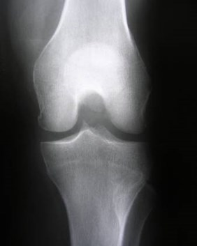
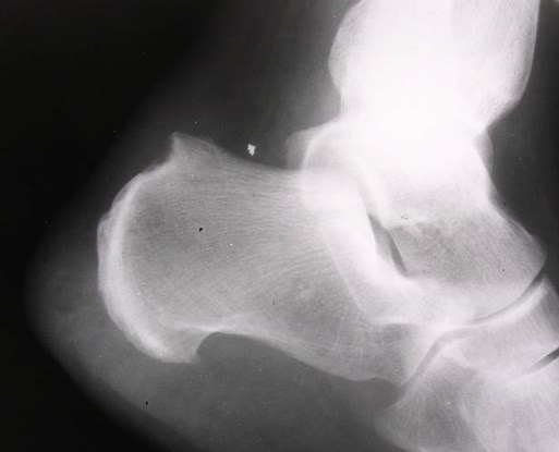
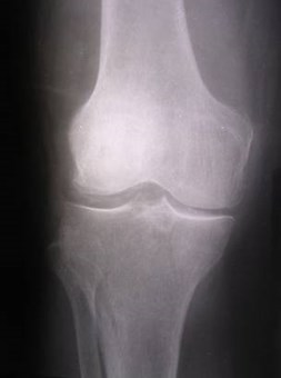
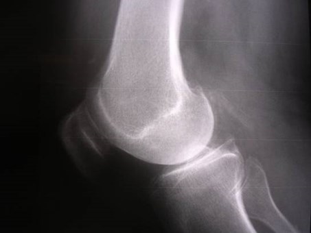
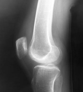
Osteoarthritis
Superior joint space narrowing,osteophytes, subchondral cysts
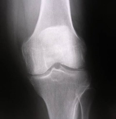
Normal opposite side
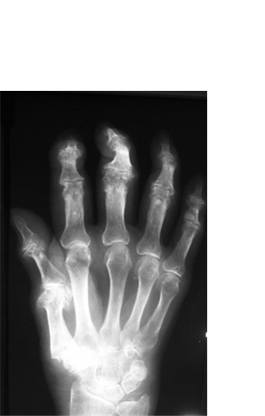
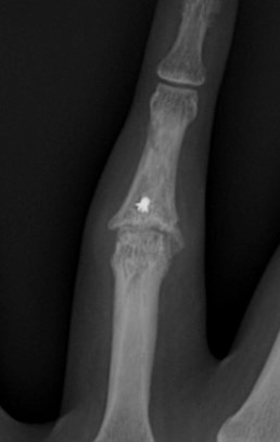
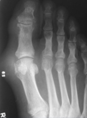
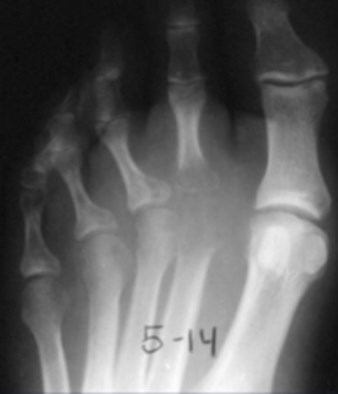
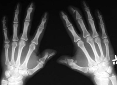
Osteoarthritis — IP joint involvement
Secondary Osteoarthritis
Another process destroys articular cartilage:
Infection
Rheumatoid arthritis
CPPD
AVN
Trauma
Hemophilia
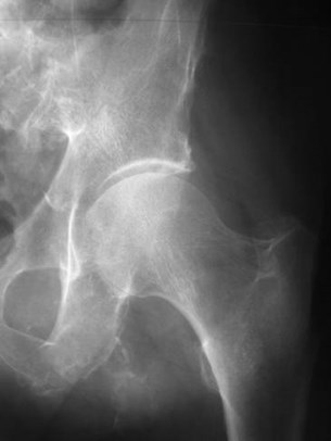
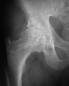
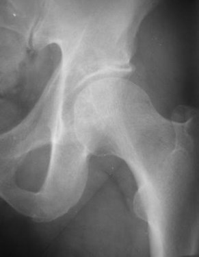
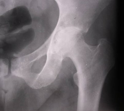
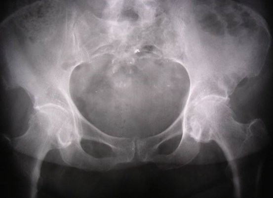
Secondary OA — primary AVN
Secondary OA – post traumatic
Inflammatory arthritis — hallmarks
Periarticular erosions
Periarticular demineralization
From regional hyperemia
Joint space narrowing
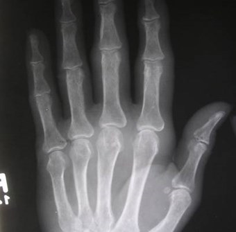
Rheumatoid Arthritis
Inflammatory arthritis– begins as synovitis
Synovial joints
Bursae
Tendons
Cartilaginous joints, ligamentous attachments notcommonly involved.
Small joints
Hands, wrists, feet
Large joints
Hips, knees, shoulders
Symmetric polyarticular disease
Rheumatoid arthritis:Progression of disease
Rheumatoid arthritis:Progression of disease
Rheumatoid Arthritis
Pathological event
Hypervascularity
Synovitis and effusion
Pannus attacks “bare”bone
Pannus attackscartilage
Radiologic findings
Periarticulardemineralization
Soft tissue swelling
Periarticular“marginal” erosions
Joint space narrowing
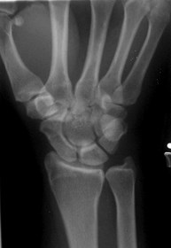
Rheumatoid arthritis
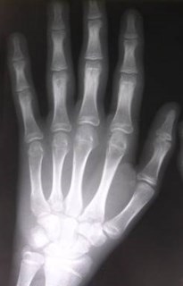
Periarticular demineralization and synovitis
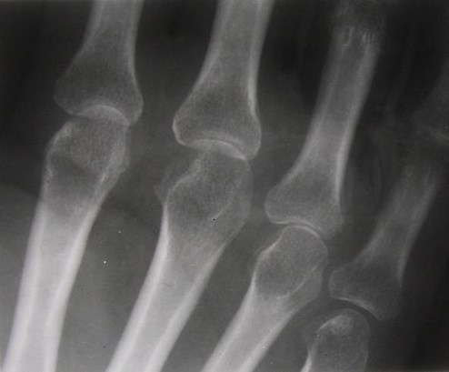
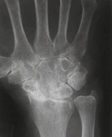
Rheumatoid arthritis
Joint space narrowing and early erosion
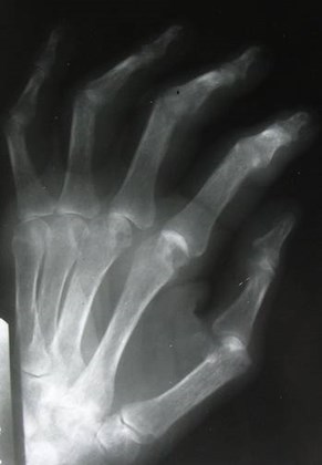
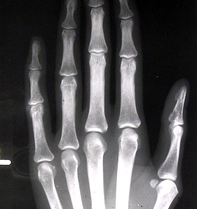
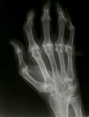
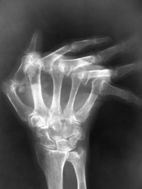
Rheumatoid arthritis
Moderate MCP erosions
Five years earlier
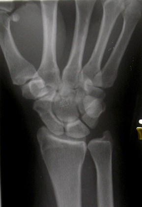
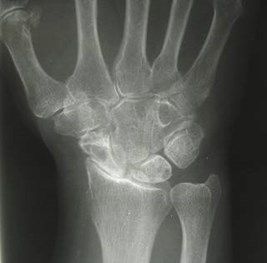
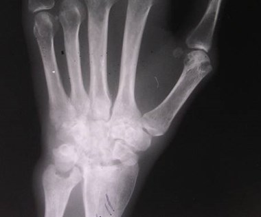
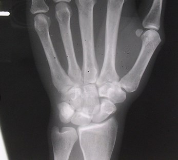
Rheumatoid arthritis
Carpal collapse
Rheumatoid arthritis
Ankylosis of wrist, PIPJ’s
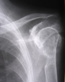
Rheumatoid arthritisLarge joints
Concentric joint space narrowing
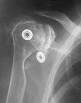
Rheumatoid arthritisLarge joints
Shoulder erosion and dislocation
Rheumatoid arthritisother manifestations
Tendon and bursa involvement
Non-articular synovitis
Bony erosion
Tendon rupture
Rotator cuff, Achilles, quadriceps tendons
Rheumatoid arthritisBursal involvement
Rheumatoid arthritis
Subacromial bursa involvement witherosion of underside of clavicle
Rheumatoid arthritisother manifestations
Subluxations andDeformities
Ulnar deviation at MCP’s
Boutonniere and Swanneck deformities offingers
Mallet finger
Almost always withtypical intra-articulardisease
Rheumatoid nodules
Subcutaneous
Proximal ulna
Achilles tendon region
Lateral fingers
Rarely calcified (DDx gouty tophi)
Almost always seropositive RA
Pathologically nonspecific
Rheumatoid nodules
Other Inflammatory Arthritis
Gout
CPPD deposition disease
Seronegitive spondylarthropathies
Reiter’s
Psoriatic arthritis
Ankylosing spondylitis
Gout
Primary or secondary
Primary gout much more common in men
Initial attack most common in 5th decade
Hyperuricemia
Lower extremities > upper
Spine, hip, shoulder disease unusual
1st MTP involved in large majority (podagra)
Limited number of joints affected, asymmetric
Gout
Acute arthritis
May simulate septic arthritis
Acute synovitis with urate crystalsin joint fluid
Strong negative birefringence
Usually no radiographic findingsexcept swelling and effusion
Gout
Chronic tophaceous gout
Crystals in cartilage, subchondral bone,synovium, periarticular tissues
Pannus formation like RA
Marginal erosions may be large, withoverhanging edges
Soft tissue masses (tophi) with calcification
Extra-articular erosions
It takes years and advanced disease toget radiographic findings
Gout
Pseudogout
Acute inflammatory arthritis due to non-urate crystals
Usually CPPD (Calcium pyrophosphatedihydrate) Deposition Disease
Chondrocalcinosis is asymptomaticprecursor
Pseudogout
effusion
chondrocalcinosis
Chondrocalcinosis
Calcification in hyaline and fibrocartilage
Menisci and articular cartilage at knee
Symphysis pubis
Triangular fibrocartilage at wrist
Articular cartilage and labrum of hip
Usually due to CPPD
Middle aged and elderly men and women
May lead to destructive arthritis similar to OA
Unusual distribution e.g. shoulder or wrist
Chondrocalcinosis
Chondrocalcinosis
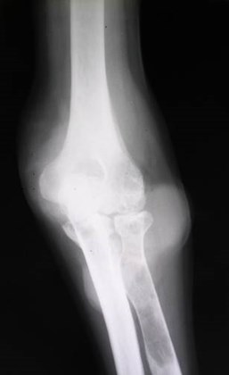
Septic arthritis
Acute
Staphlococcal
Gonococcal
Chronic
Tuberculous
Fungal
Hematogenous
Large joints
Direct extension
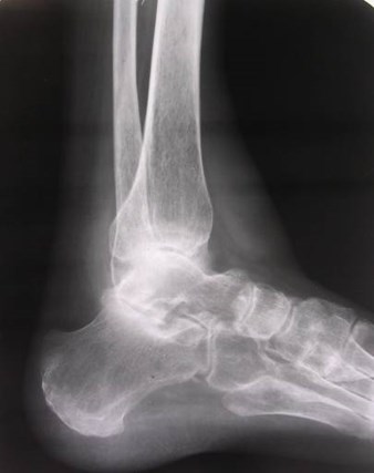
Diabetic foot with ulcer
Septic arthritis
Early
Severe pain and limitation of motion
Effusion, soft tissue swelling
Later
Joint space narrowing
Subchondral bone destruction
Bony findings indicate severe damage tocartilage
Early intervention is crucial
Don’t wait for radiographic findings!
Diabetic foot with swelling
Diffuseperiarticulardemineralizationfrom hyperemia
Articularsurfaces andsubchondralbonedestroyed
Joint effusion
If acute symptoms, think infection, goutor pseudogout, trauma or hemorrhage
Seronegative Spondyloarthropathies
“Rheumatoid variants”
Psoriatic arthritis
Reiter’s syndrome
Ankylosing spondylitis
Inflammatory bowel disease
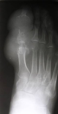
Psoriatic arthritis
Asymmetric
Spine
Large asymmetric osteophytes
Fingers
DIPJ
PIPJ
Severely erosive arthritis
“pencil in cup” deformity
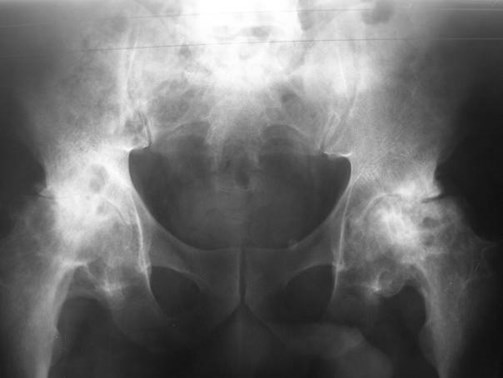
Sacroiliitis
asymmetric
Reiter’s syndrome
Classic triad
Arthritis (50%)
Urethritis
Uveitis
Reiter’s syndrome
Asymmetric disease (unlike RA)
Foot > hand
Hip and knee
Sacroiliitis
Regional osteoporosis - maymimic RA
Enthesopathy – whiskering attendinous insertions
Proliferative plantar heel spur
Large asymmetric spinalosteophytes
Review Questions
Periarticular erosions are not acommon feature of:
Rheumatoid arthritis
Osteoarthritis
Gout
Psoriatic arthritis
Osteophytes can be seen in:
Osteoarthritis
Psoriatic arthritis
Post-traumatic arthritis
Avascular necrosis
All of the above
Septic arthritis: True or false?
The earliest radiographic sign is jointeffusion.
Joint space narrowing is an early sign.
Imaging should not be the first step inevaluation of suspected joint infection.
Joint surface erosion indicates severedisease.
Additional reading
1.Resnick, D Target Approach to Articular Disorders,Chapter 46 in Bone and Joint Disorders 2nd editionW.B. Saunders Co. Philadelphia 1996.
2.Resnick, D (see above) Chapter 22 RheumatoidArthritis.
3.Greenspan, A Degenerative Joint Disease,Chapter 10 in Orthopedic Radiology: A PracticalApproach JB Lippincott Co. Philadelphia 1988
4.Jones AC et al. Diseases associated with calciumpyrophosphate deposition disease. Semin ArthritisRheum 1992; 22:188.

The End
Use the back button on the browser to exit the program