|
Spondylolesthesis
· Normally the inferior articulating fact of each lumbar vertebral body lies posterior to the superior articulating facet of the body below it
· If the bony connection between the inferior and superior articulating facets (pars interarticularis) is defective, the weight of the body may cause the upper vertebra, including all of the vertebral bodies above it, to slip forward for varying amounts on the body below (spondylolisthesis)
· The defect in the pars interarticularis is called spondylolysis
· Spondylolysis is not present at birth but increases in frequency with increasing age
· When symptomatic, pain is the most frequent symptom of either spondylolysis or spondylolisthesis
· There are several different etiologies for spondylolisthesis
o Spondylolytic spondylolisthesis
§ Is the most common type and results from bilateral defects in the pars interarticularis
§ Has a 2:1 male to female predominance and is more common in Caucasians than African-Americans
§ With bilateral spondylolysis, the posterior aspect of vertebral body separates from the anterior body
· Posterior body remains fixed in position while the anterior part is free to slip forward
§ Cause of spondylolysis appears to be a combination of a dysplastic pars at birth coupled with the long-term stresses of upright posture
§ Certain athletic endeavors (e.g., football, weight-lifting, tennis and wrestling) appear to increase these stresses
§ Fractures of the pars may heal with a pseudarthrosis or fibrous ankylosis
o Degenerative spondylolisthesis
§ More common in African American women than in Caucasian women
§ Most frequent at the L4-L5 level
§ Is not the result of a pars defect but a complex interaction between the disk, facets joints and the ligamentous structures
§ There is usually narrowing, sclerosis of the facet joints from osteoarthritis
§ There is less forward slippage (spondylolisthesis) in this group than in spondylolytic group
§ Retrolisthesis (backward slippage of a vertebral body on the body below it) may occur with osteoarthritis of the facets joints
o Dysplastic spondylolisthesis
§ Results from congenital abnormalities of the body and/or facets in the lumbar region such that the alignment of the facets allows spondylolisthesis to occur
§ The pars may or may not be intact
§ Females are more apt to have this type than males (2:1)
§ Symptoms usually develop during the adolescent growth period
o Traumatic spondylolisthesis
§ Trauma can lead to an acute fracture through a normal pars interarticularis
§ Results in a diastatic defect that may lead to a spondylolisthesis
· Imaging findings
o Conventional radiography in the anteroposterior, lateral and both oblique projections is usually adequate to demonstrate both spondylolysis and spondylolisthesis
o Spondylolysis appears as a break in the “neck” of the Scottie Dog on the oblique view
o Bilateral spondylolysis is visible on the lateral view
o Forward slippage of one vertebral body on the other is observed and graded on the lateral view
o Spondylolisthesis is graded in this manner
§ Grade 1-vertebral body above subtends ¼ of the AP diameter of the vertebral body below
§ Grade 2- vertebral body above subtends 1/2 of the AP diameter of the vertebral body below
§ Grade 3- vertebral body above subtends 3/4 of the AP diameter of the vertebral body below
§ Grade 4- vertebral body above subtends the full AP diameter of the vertebral body below

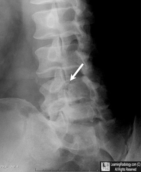
Spondylolytic Spondylolisthesis. Lateral view of the lumbar spine demonstrates
a bilateral break in the pars interarticularis or spondylolysis (lucency shown by black arrow)
that allows the L5 vertebral body (red arrow) to slip forward on the S1 vertebral body (blue arrow).
The forward slippage is called spondylolisthesis. The normal pars interarticularis is shown by the white arrow. The degree of forward slippage is equal to about 1/4 to 1/2 of the AP diameter of S1
so this is a Grade1-Grade 2 spondylolisthesis.
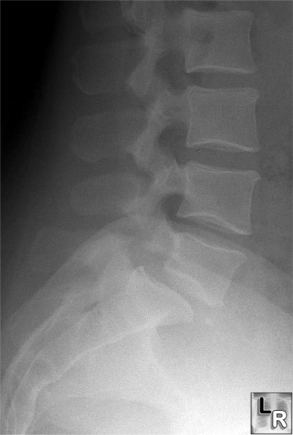
For a photo of the same image without arrows, click here
Spondylolysis. The white arrow points to a "break" in the "neck" of the Scottie Dog representing spondylolysis at L4 involving the right pars interarticularis.
|
