|
|
Hyperostosis Frontalis Interna
General Considerations
- Occurs mostly in post-menopausal females over 35 years of age
- Thickening of the inner table of the skull
- Unknown etiology although its presence in post-menopausal women suggest possible hormonal influence
Clinical Findings
- No known clinical significance
- May be more prevalent with obesity
Imaging Findings
- Bilateral and symmetrical
- Cortical thickening that affects primarily frontal bone
- May occasionally extend to parietal bones
- Characteristically does not extend across the midline at the sagittal sinus
- May appear flat or nodular
Differential Diagnosis
- Should not be confused with hyperostosis from tumor or metastatic disease, Paget disease or fibrous dysplasia

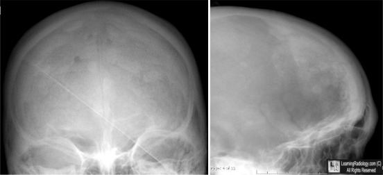
Hyperostosis frontalis interna. Frontal view of skull on left shows sclerosis in a patchy almost nodular appearance (blue arrows) which characteristically does not cross the midline (black arrow). On the lateral view on the right, the hyperostosis is confined to the frontal bone (white arrow).
For more information, click on the link if you see this icon 
For this same photo without the annotations, click here
Prevalence of hyperostosis frontalis interna in relation to body weight. Verdy M, Guimond J, Fauteux P and Aube M. The American Journal of Clinical Nutrition 31: Nov 1978, pp. 2002-2004.
|
|
|
