|
|
Cerebellar Pilocytic Astrocytoma
Submitted by Terence Menezes, MD
The patient has known neurofibromatosis, type I. There are numerous patchy foci of high signal within the cerebellum and the brainstem, and subtle change in the thalami.
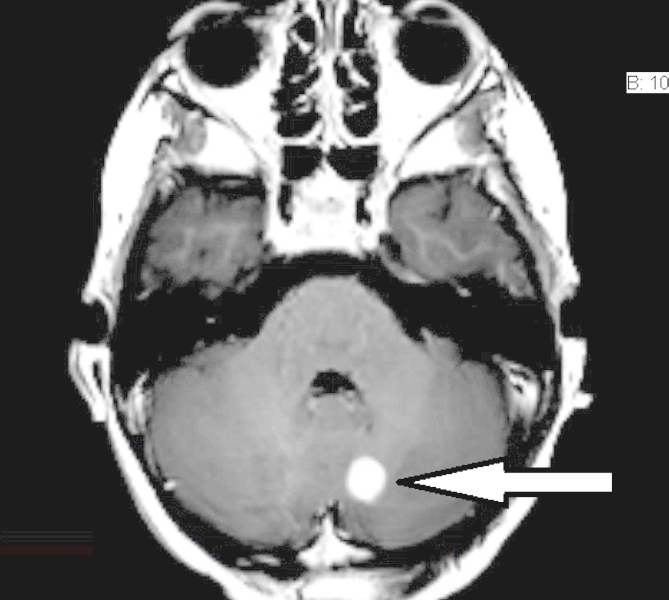
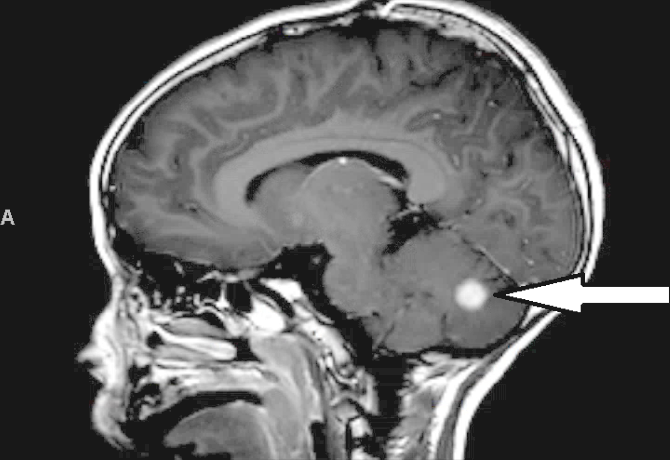
There is a very well defined, almost spherical lesion in the medial left cerebellar hemisphere; it is frankly hyperintense on T2 and FLAIR, measuring 13.8 mm AP by 11.0 mm transverse.
On contrast images in three planes, this same lesion measures 10.8 mm AP by 10.8 mm transverse by up to 9.2 mm craniocaudad, which represents an increase in size compared to the previous MRI of 07/16/2014, when it had measured 6 mm.
|
|
|
