Etiology
o
Inflammation of the renal parenchyma and renal
pelvis
due to an infectious source
o
Most often secondary to an ascending lower
urinary
tract infection from gram-negative bacteria
§
E. coli
§ Klebsiella
§ Proteus
§ Pseudomonas
o
Exception is S. aureus, which is spread hematogenously
·
Pathologic Causes
o
Vesicoureteral reflux
o
Obstruction in the collecting system usually due
to a
calculus
·
Signs and symptoms
o
Fever
o
Chills
o
Flank pain
o
Dysuria
o
Increased frequency of urination.
o
On exam, costovertebral angle tenderness may be
present.
· Clinical Findings
o
CBC
§
Elevated white blood cell count.
o
Urinalysis
§
Bacteriuria
§
Pyuria
§
White blood cell casts
o
Acute pyelonephritis is clinical diagnosis,
§ Radiographic imaging is used to evaluate
underlying
pathology
§ Rule out any complications.
· Complications
o
Abscess
o
Emphysematous pyelonephritis
§
Most often occurs in diabetics
· Can produce gas in the collecting system and
renal parenchyma
· Imaging Findings
o
Enlarged kidneys (U/S and CT)
o
Hydronephrosis (U/S and CT)
o
Wedge shaped areas of low attenuation secondary
to
decreased perfusion (CT)
o
Loss of the ability to distinguish the
corticomedullary
border (CT)
o Perinephric stranding. (CT)
·
Treatment
o
Antibiotics for non-complicated pyelonephritis.
o
Radical nephrectomy for emphysematous
pyelonephritis.
o
Percutaneous drainage of abscesses
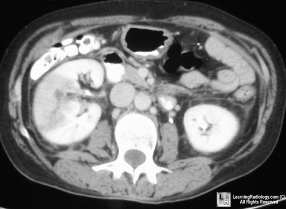
Acute Pyelonephritis. Right kidney is markedly enlarged and
has a wedge-shaped area of low attenuation.