|
|
Scleroderma of the Gastrointestinal Tract
General Considerations
- Gastrointestinal manifestations of scleroderma are relatively common (40-45%), following skin changes and Raynaud’s phenomena
- In the GI tract, there may be atrophy of the smooth muscle
- CD4 T cells are believed to play an important role in its pathogenesis
- The esophagus is affected most frequently (85%) with lesser changes in the stomach (uncommon), small (45%) and large bowel
Clinical Findings
- Many patients are asymptomatic despite significant GI changes
- Dysphagia
- Reflux
- Delayed gastric emptying
- Malabsorption
- Pseudo-obstruction
- Constipation
Imaging Findings
- Contrast-enhanced conventional radiography (i.e. barium studies) are usually used to image GI scleroderma
- Esophagus
- Dilated, hypomotile esophagus specially involving distal 2/3
- Patulous esophagogastric junction
- Delayed emptying of esophagus in recumbent position but improved emptying when upright (DDx: achalasia)
- Distal esophageal stricture from reflux esophagitis
- Barrett esophagus
- Adenocarcinoma from Barrett esophagus
- Stomach
- Involvement is rare but there may be dilatation and hypomotility
- Small bowel
- Dilation, especially duodenum and jejunum
- Fragmentation and flocculation of barium
- “Hide-bound” appearance with valvulae close together despite small bowel dilatation
- Pseudo-diverticula on the mesenteric side
- Colon
- Large-mouth pseudo-diverticula on anti-mesenteric border, usually in transverse and descending colons
- Dilatation
- Effacement of the haustra
Differential Diagnosis
- Achalasia
- Small bowel obstruction
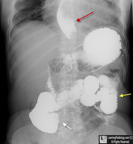
Gastrointestinal Scleroderma. A single view of the lower chest and abdomen after an upper gastrointestinal barium study demonstrates a dilated, distal esophagus with reflux (red arrow, a normal-appearing stomach and dilation of the small bowel (white arrow) with relatively close approximation of the valvulae-the "hide-bound appearance" (yellow arrow).
Gastrointestinal Scleroderma Imaging. AN Khan and R Rahim. eMedicine
|
|
|
