|
|
Feline Esophagus
"Esophageal Shiver"
General Considerations
- Thin, horizontal folds that traverse the entire lumen of esophagus
- Usually seen in distal 2/3 of esophagus
- So-named because of their resemblance to the appearance of the distal esophagus in cats
- Transient phenomenon seen during double-contrast esophagrams
- Most frequently observed in patients with gastroesophageal reflux and hiatal hernia
Differential Diagnosis
- Thicker, transverse folds which do not extend across the entire lumen seen with reflux esophagitis scarring
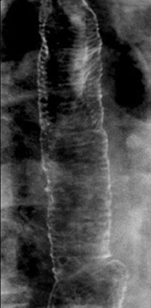
Feline Esophagus. There are multiple, thin, transverse lines seen crossing the entire esophageal lumen on this double-contrast esophagram.
Feline Esophagus. EE Furth, SE Rubesin and, D Rose. AJR 1995;164:90
|
|
|
