|
|
Paget Disease
General Considerations
- Multifocal chronic skeletal disease due to chronic paramyxoviral infection
- Prevalence
- 3% of individuals >40 years
- 10% of persons >80 years
- Unusual <40 years
- M:F = 2:1
- Active or Osteolytic phase
- Aggressive bone resorption with lytic lesions
- Replacement of hematopoietic bone marrow by fibrous connective tissue with numerous large vascular channels
- Inactive or Quiescent phase
- Decreased bone turnover with skeletal sclerosis and thickening of the cortex
- Mixed pattern
- Lytic and sclerotic phases frequently coexist
Clinical findings
- Asymptomatic (1/5)
- When symptomatic, symptoms may include
- Fatigue
- Enlarged hat size
- Peripheral nerve compression
- Neurologic disorders from compression of brainstem (basilar invagination)
- Hearing loss, blindness
Imaging Findings
- Classical triad
- Thickening of the cortex
- Accentuation of the trabecular pattern
- Increased size of bone
- Cyst-like areas
- Skull (involvement in 29-65%)
- Inner and outer table involved
- Leads to diploic widening
- Osteoporosis circumscripta is well-defined lysis, most commonly in frontal bone producing well-defined geographic lytic lesion in skull
- Represents early destructive phase of disease active stage)
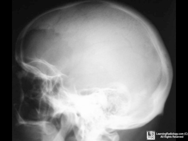
Osteoporosis circumscripta of frontal bone
in lytic phase of Paget's disease
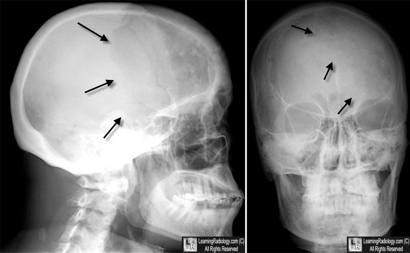
Osteoporosis circumscripta of frontal bone
in lytic phase of Paget's disease (black arrows) on the lateral and frontal projection.
- "Cotton wool" appearance represents mixed lytic and blastic pattern of thickened calvarium (later stage)
- Basilar invagination with encroachment on foramen magnum
- Deossification and sclerosis in maxilla
- Sclerosis of skull base
- Long bones (almost invariably starts at end of bone)
- "Candle flame" or "blade of grass" pattern of lysis is the advancing tip of V-shaped lytic defect in diaphysis of long bone originating in subarticular site
- Lateral curvature of femur
- Anterior curvature of tibia (commonly resulting in fracture)
- Pelvis

- Thickened trabeculae in sacrum, ilium
- Rarefaction in central portion of ilium (looks like a large lytic lesion)
- Thickening of iliopectineal line
- Acetabular protrusio with secondary degenerative joint disease
- Spine (upper cervical, low dorsal, midlumbar most common sites)
- Coarse trabeculations at periphery of bone
- "Picture-frame vertebra" mimics bone-within-bone appearance
- Enlarged vertebral body with reinforced peripheral trabeculae and more lucent center, typically in lumbar spine
- "Ivory vertebra" is a blastic vertebra with increased density
- Ossification of spinal ligaments, paravertebral soft tissue, disk spaces can occur
DDx
- Depends on the bone in which it occurs
- Skull
- Osteolytic or osteoblastic metastases
- Long bones
- Metastases
- Chronic osteomyelitis (thickened cortex)
- Old trauma (thickened cortex)
- Hodgkin’s disease
- Spine
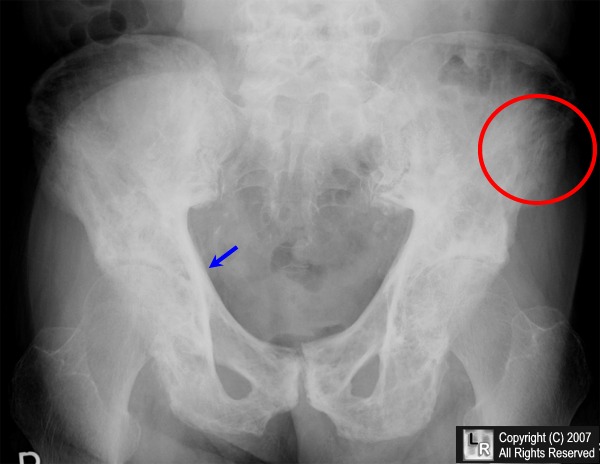
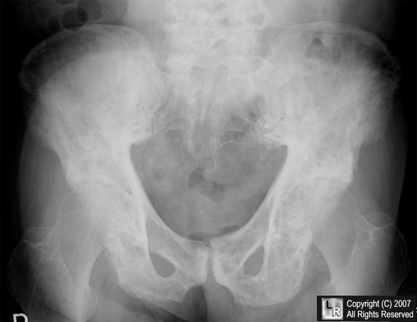
Paget's Disease -- pelvis. Frontal radiograph of the pelvis demonstrates the classical triad of thickening
of the cortex (blue arrow), accentuation of the trabecular pattern (red circle) and increased density of the bone.
For additional information about this disease, click on this icon above.
For this same photo without the arrows, click here
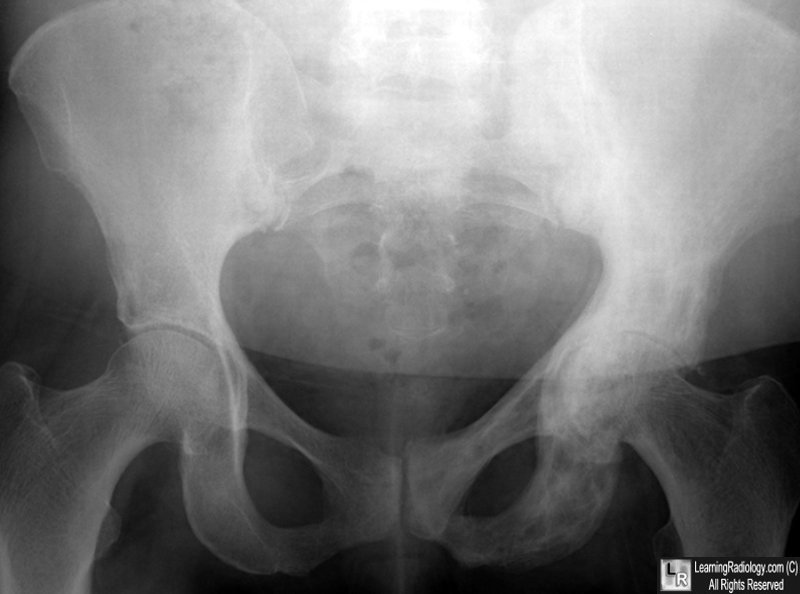
Paget Disease, Left Hemipelvis. The entire left hemipelvis demonstrates increased bone density with thickening of the cortex and accentuation of the trabecular markings characteristic of paget Disease.
|
|
|

)