|
|
Pneumonia - Right Lower Lobe
General Considerations
- It is always best to localize disease on conventional radiographs
using two views taken at 90° to each other (orthogonal views) like
a frontal and lateral chest radiograph
- Sometimes, only a frontal radiograph may be available, as in
critically ill or debilitated patients who require a portable
bedside examination
- Nevertheless, it is still frequently possible to localize the
pneumonia
using only the frontal radiograph by analyzing which
structure’s
edges are obscured by the disease
Air Bronchogram
- Pneumonia may contain air bronchograms if the bronchi themselves
are not filled with inflammatory exudate or fluid
- When the bronchi are filled with fluid, as in bronchopneumonia,
there will be no air bronchograms present
- Air bronchograms are much more likely to be visible when the
pneumonia involves the central portion of the lung near the hilum
- Near the periphery of the lung, the bronchi are usually too small
to be visible
- Remember that anything of fluid or soft tissue density that replaces
the normal gas in the airspaces may also produce this sign so an air
bronchogram is not specific for pneumonia
Lobar Pneumonia
- The prototypical lobar pneumonia is pneumococcal pneumonia
caused by Streptococcus pneumoniae
- Although we are calling it lobar pneumonia, the patient may present with the disease before the entire lobe is involved
- In its most classical form, the disease fills most or all of a lobe
of the lung
- Since lobes are bounded by interlobar fissures, one or more of
the margins of a lobar pneumonia may be sharply marginated Where the disease is not bound by a fissure, it will have an indistinct
and irregular margin
- Lobar pneumonias almost always produce a silhouette sign where they come in contact with the heart, aorta or diaphragm and they
almost always contain air bronchograms if they involve the central
portions of the lung
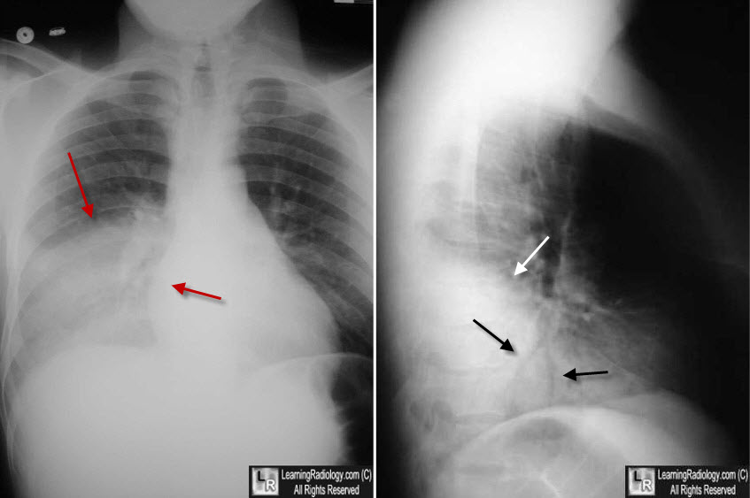
Right Lower Lobe Pneumonia. The frontal view shows an airspace density in the right lower lung
field (red arrows) that has a distribution corresponding to the location of the right lower lobe. The lateral view
confirms the pneumonia is posterior (white arrow), and contains two, black-branching structures that are air bronchograms (black arrows),
|
|
|
