|
|
Pulmonary Laceration
Traumatic Pneumatocele
General Considerations
- Most thoracic trauma is due to motor vehicle accidents
- Most thoracic trauma is blunt, rather than penetrating
- Lacerations usually result from blunt chest trauma
- They represent tears in the lung parenchyma
- Lacerations filled with air are called pneumatoceles, and those filled with blood are called pulmonary hematomas
- They tend to occur more often in children and young adults
Clinical Findings
- No symptoms from laceration itself unless it becomes infected,
which is rare
Imaging Findings
- Usually not apparent at first because of surrounding pulmonary
contusion
- Contusions characteristically clear rapidly, sometimes within
48 hours
- On CT, they will present as cystic lucencies, frequently beneath a rib fracture
- CT is more sensitive than conventional radiographs for detecting a pulmonary laceration
- Half are solid, mass-like lesions (pulmonary hematoma)
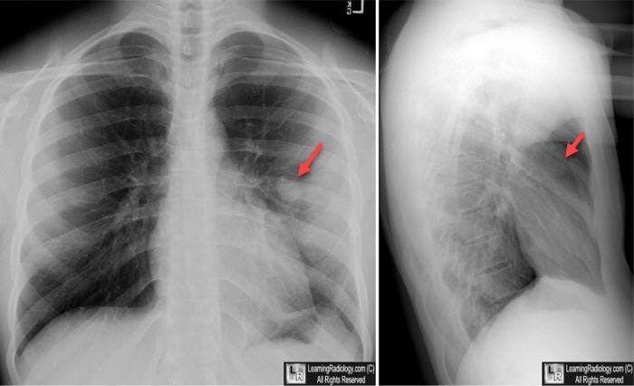
Pulmonary Hematoma. Following blunt chest trauma on the left, a small, solid-appearing, soft tissue nodule appeared in the lingula (red arrows) representing pulmonary laceration filled with blood.
- Half are thin-walled cystic structures (traumatic lung cyst
or pneumatoceles) with or without air-fluid level — depends on
how much bleeding into laceration
- Usually subpleural location under point of maximum impact
- May be single or multiple
Treatment
- Supportive: oxygen, assisted ventilation if needed
Prognosis
- Characteristically, they take a long time to heal – weeks to months
- Gradually decrease in size
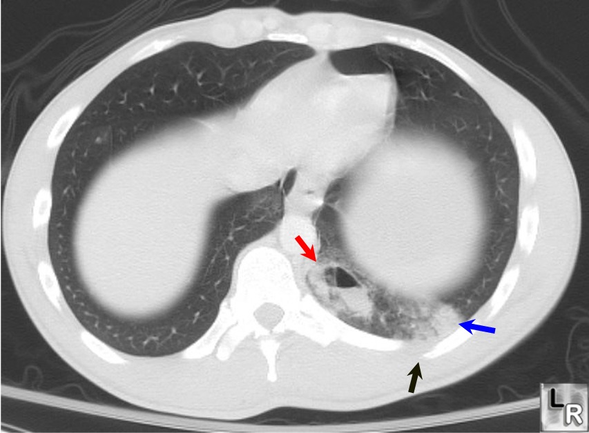
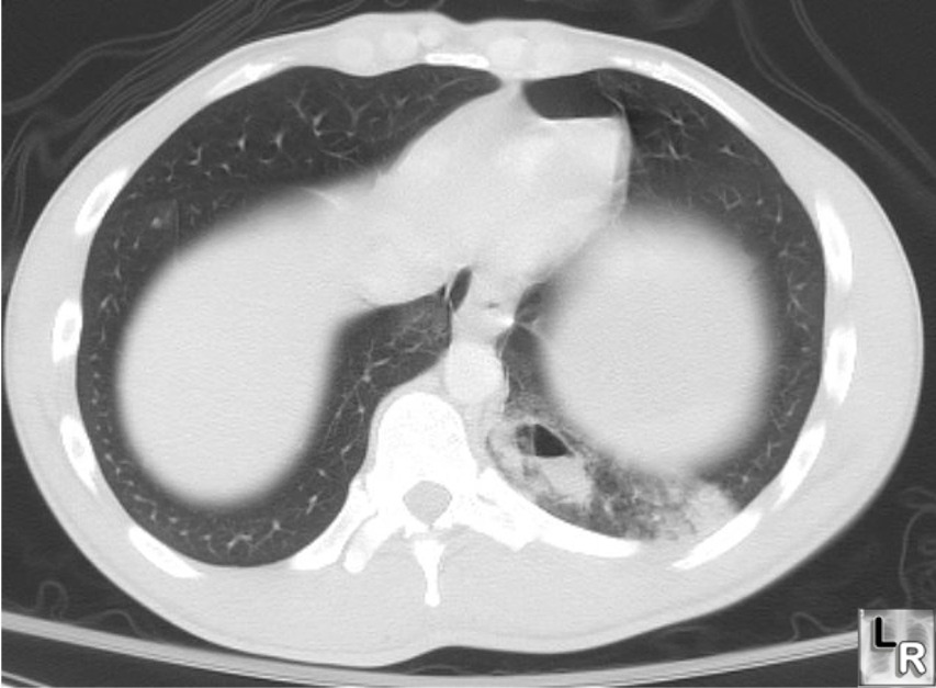
Pulmonary laceration. CT of the chest demonstrates a partially-cystic,
partially fluid-filled structure in the left lower lobe (red arrow) near a rib fracture (black arrow)
in this patient who was an unrestrained passenger in a motor vehicle collision. The blue arrow
points to an area of subpleural hemorrhage representing a pulmonary contusion
For a photo of the same image without arrows, click here
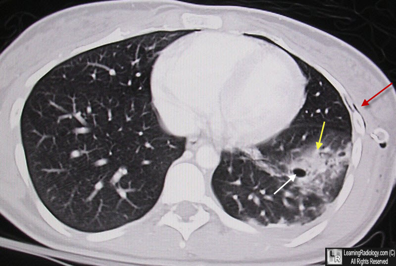
Pulmonary laceration. CT of the chest again demonstrates a cystic,
partially fluid-filled structure in the left lower lobe (white arrow) surrounded by a pulmonary contusion (yellow arrow) There is subcutaneous emphysema (red arrow) from the stab would.
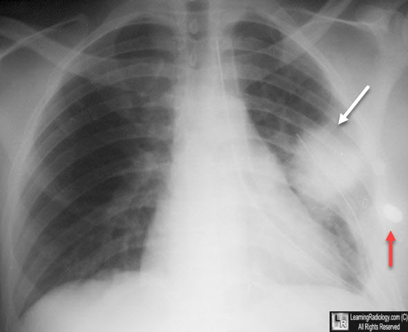
Pulmonary laceration. Upright chest radiograph shows a "solid"
appearing structure in the left hemithorax representing a blood-filled traumatic pneumatocoele. The patient had been shot (red arrow points to bullet). There are already two chest tube in place.
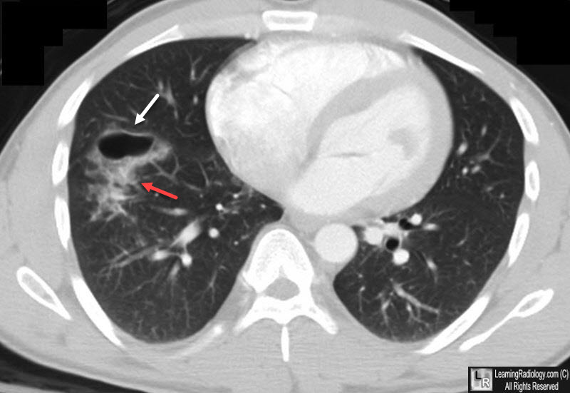
Pulmonary laceration. Chest CT shows a thin-walled cavity (white arrow) with
surrounding pulmonary hemorrhage (red arrow).
|
|
|
