|
|
Pneumopericardium
General Considerations
- Air in the pericardial space
- Causes include:
- Penetrating trauma (rarely, blunt chest trauma)
- Assisted ventilation with severe lung pathology, especially in infants but also asthmatics (barotrauma)
- Thoracic surgery which violates the pericardial sac
- In adults, pneumopericardium is much less common than pneumomediastinum, the former usually not occurring in adults unless there is a direct injury to the pericardium
Clinical Findings
- Depend on the amount of air and how rapidly it accumulates
- Substernal chest pain
- Hypotension, bradycardia, hypoxia
- Hamman’s sign: a continuous crunching or clicking noise synchronous with the heart beat heard best in the left lateral decubitus position
Imaging Findings
- The heart is partially or completed surrounded by a rim or air-density
- The parietal pericardium may be visible as a thin line
- Characteristically, air in the pericardium (unlike air in the mediastinum) remains below the level of the great vessels
Differential Diagnosis
- Pneumomediastinum (see above)
Treatment
- Can be self-limited and resolve spontaneously
- Cardiac tamponade can occur, however, and aspiration or pericardial drainage tube can be used
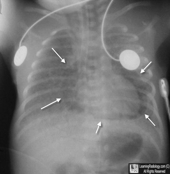
Pneumopericardium. There is marked "ground-glass" opacification to both lungs in this newborn with severe hyaline membrane disease on assisted ventilation. Surrounding the heart is a band of air-density (white arrows) which does not extend above the level of the root of the aorta or pulmonary arteries, characteristic of pneumopericardium. Incidentally, the tip of the endotracheal tube is down too far as it extends into the right main bronchus.
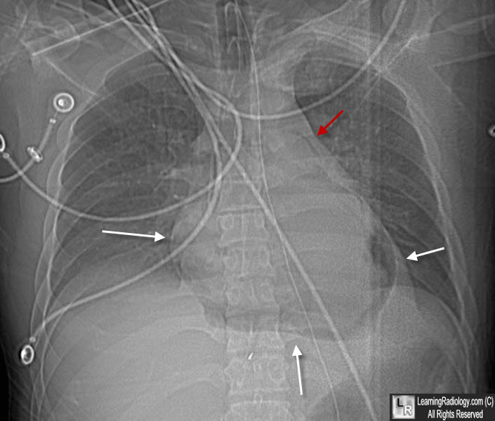
Pneumopericardium. This is the "scout" image from a CT scan of the chest. Notice the halo of air surrounding the heart (white arrows) that does not extended above the level of the great vessels (red arrow). The patient was stabbed in the chest.
Stacey S et al. A case of spontaneous tension pneumopericardium. Br J Cardiol 2004;11:32-14.
|
|
|
