|
|
Pneumonia - Left Upper Lobe
General Considerations
- It is always best to localize disease on conventional radiographs using two views taken at 90° to each other (orthogonal views) like a frontal and lateral chest radiograph
- Sometimes, only a frontal radiograph may be available, as in critically ill or debilitated patients who require a portable bedside examination
- Nevertheless, it is still frequently possible to localize the pneumonia using only the frontal radiograph by analyzing which structure’s edges are obscured by the disease
Airspace disease may contain air bronchograms
- The visibility of air in the bronchus because of surrounding airspace disease is called an air bronchogram
- An air bronchogram is a sign of airspace disease
- Bronchi are normally not visible because their walls are very thin, they contain air and they are surrounded by air
- When something like fluid or soft tissue replaces the air normally surrounding the bronchus, then the air inside of the bronchus becomes visible as a series of black, branching tubular structures—this is the air bronchogram
- What can fill the airspaces besides air?
-
Fluid, such as occurs in pulmonary edema
-
Blood, e.g., pulmonary hemorrhage
-
Gastric juices, e.g., aspiration
-
Inflammatory exudate, e.g., pneumonia
-
Water, e.g., near-drowning
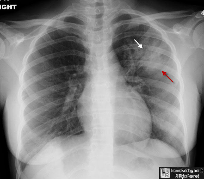
Left Upper Lobe Pneumonia. There is airspace disease in the left upper lobe with fluffy, indistinct margins (red arrow) containing air bronchograms (white arrows). This was found to be a staphylococcal pneumonia.
|
|
|
