|
|
Incomplete Rim Sign
General Considerations
- The concept, seen mainly on conventional radiographic images, that a pedunculated “mass” may display a portion of its border that is indistinct where the “mass” is attached and another portion of the border which is sharply marginated where the structure projects into air
- The finding helps to establish that a “mass” has a soft tissue attachment and projects into an air-filled density
- That is, the “mass” is not completely surrounded by air
- Examples may include
- The normal nipple shadow
- A mass attached to the chest wall and projecting out into the atmosphere (e.g., a mole or keloid)
- A mass attached to the pleural surface and projecting into the air-filled lung (e.g. a pleural-based mass)
- An abdominal hernia that is attached to the body and projects outward into the air-filled atmosphere
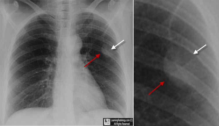
Incomplete Rim Sign. A mass, in this case an extrapleural lipoma, demonstrates one border which is sharply demarcated (red arrows) where it projects into the air-filled lung, and another, indistinct border (white arrows) where it is attached to the chest wall.
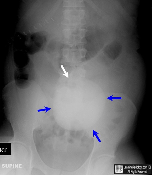
Incomplete Rim Sign. An umbilical hernia demonstrates a sharp border (blue arrows) where it projects outward into the air-filled atmosphere, and an indistinct border (white arrow) where it is attached to the body.
|
|
|
