|
|
Hydropneumothorax
General Considerations
- Abnormal accumulations of fluid along with air in the pleural space
Causes
- Trauma
- Thoracentesis
- Prior surgery
- Bronchopleural fistula
Imaging Findings
- On conventional radiography done with the x-ray beam directed horizontally (typically with the patient upright), there will be an air-fluid level in the thoracic cavity
- Pleural fluid without a pneumothorax characteristically produces a meniscus-shaped upper border to its density
- On supine radiographs, a hydropneumothorax will be more difficult to see although a uniform grayness to the entire hemithorax with the absence of vascular markings suggest the diagnosis
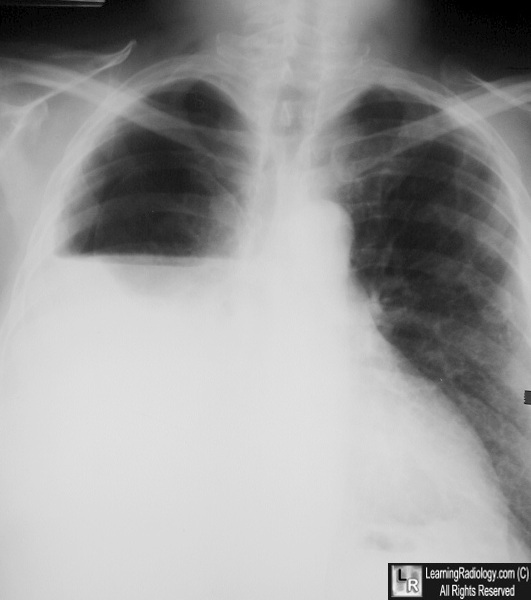
Hydropneumothorax. There is an air-fluid level in the right hemithorax on this upright chest radiograph because there is both fluid (in this case, blood) and air in the right pleural space. The patient was stabbed.
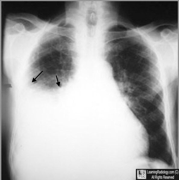
Right pleural effusion. There is a moderately large right pleural effusion present. As there is no pneumothorax, the effusion produces a characteristic meniscus shape, slightly higher laterally and medially than in the center (black arrows).
|
|
|
