|
|
Dense Hilum Sign
General Considerations
- Appearance of the hilar shadow on the frontal chest radiograph in which the hilar shadow on the affected side appears denser than its counterpart on the opposite side
- This may be due to a mass or calcification in the hilum but, if the hilum is not enlarged or calcified, then the sign can also be seen when there is the superimposition of another abnormal density in the lung or mediastinum that projects over the hilum on the frontal image
- This can frequently be seen with pneumonia or a mass that occupies the superior segment of either lower lobe, a segment that will superimpose on the hilum in the frontal projection
- The lateral projection is necessary for evaluating the cause of a dense hilum
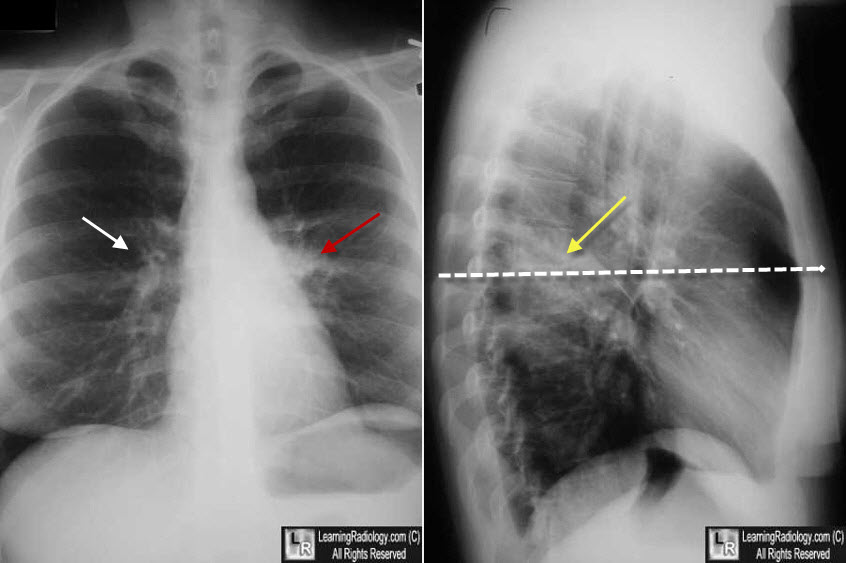
Dense Hilum Sign. On the frontal (PA) image, the left hilum (red arrow) appears denser than the right hilum (white arrow). This may be caused by a hilar mass, but not necessarily. The lateral view shows airspace disease (pneumonia) in the superior segment of the left lower lobe (yellow arrow). The hilum appears dense on the frontal image because the x-ray beam (dotted white arrow)passes through BOTH the hilum and superior segment on the PA projection, causing them to superimpose on one another.
|
|
|
