|
|
Aspergilloma
- Histologically, these are intertwined
hyphae of the aspergillus
forming a
mycetoma
-
Solid, round mass in thin-walled cavity
-
Usually in upper lobes
-
> Moves with changes in positioning
-
Crescent-shaped airspace separates
the fungus ball from the wall of cavity
-
Fungus ball may calcify

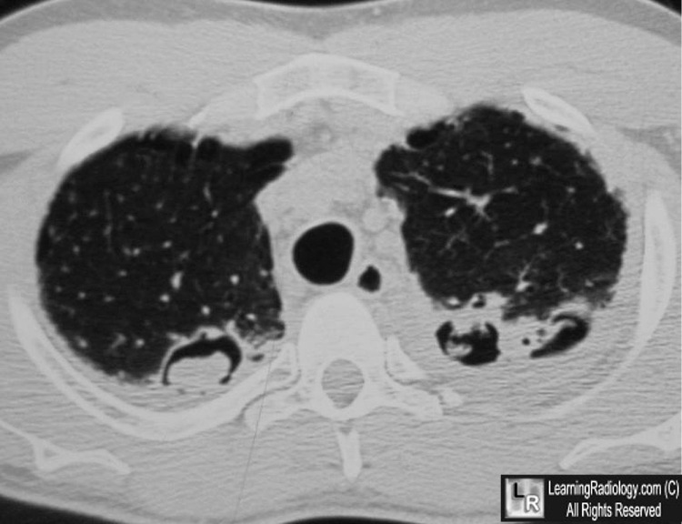
Aspergillomas, both upper lobes. White arrows point to two masses contained within cavities
in both upper lobes, the one on the right larger than the left. The masses represent aspergillomas
in cavities of a patient with a prior history of sarcoidosis.
For this same photo without arrows, click here
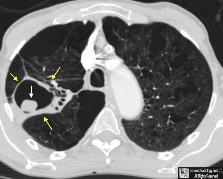
Aspergilloma. A nearly-round fungus ball (white arrow) has formed in a prior tuberculous cavity (yellow arrows), The patient is supine so the aspergilloma has moved to the dependent portion of the cavity in which it formed.
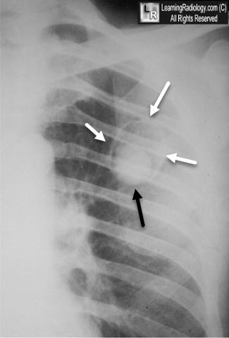
Aspergilloma. There is a thin-walled upper lobe cavity (white arrows) presumably from old TB, with a fungus ball in the dependent portion (black arrow).
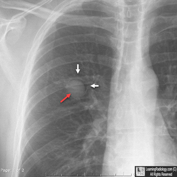
Aspergilloma. There is a thin-walled upper lobe cavity (white arrows) presumably from old TB, with a fungus ball in the dependent portion (red arrow).
For more information, click on the link if you see this icon 
|
|
|
