|
|
Acute Chest Syndrome
General Considerations
- Leading cause of death in patients with sickle cell disease
- Syndrome is characterized by
- New airspace disease on chest x-ray, and one or more of the following:
- Fever (variable)
- Cough
- Sputum production
- Shortness of breath
- Hypoxia
- Exact cause is unknown
- Most often affects those with homozygous sickle cell and sickle cell-beta thalassemia genotypes
- More common in the winter, in younger individuals, and after surgery
- More severe in adolescents and adults (>20) than in children
Clinical Findings
- May be recurrent
- Clinical course is usually rapid
- Symptoms can range from mild to fatal
- Blood and sputum cultures are frequently negative
- There may be chest wall tenderness from rib infarctions
Imaging Findings
- Pathogenesis of all of the airspace disease in ACS is not fully understood
- Part of it is commonly caused by non-embolic thrombosis of sickled erythrocytes in the pulmonary vasculature
- Radiographs may be normal at the start of an episode
- On conventional radiographs
- Patchy airspace disease in a segmental, lobar or multilobar distribution
- May or may not have a pleural effusion
- Airspace disease may also be made up of atelectasis, pneumonia or fat embolism
- Fat embolism occurs from bone marrow necrosis and is thought by some to play a key role in the pathogenesis of the syndrome
- On CT, there may be ground-glass opacities in a patchy, mosaic or multifocal pattern of distribution
Differential Diagnosis
- Pulmonary edema
- Pulmonary hemorrhage
Treatment
- Antibiotics
- Fluids
- Supplemental oxygen
- Bronchodilators
- Pain medication
- Transfusions
- Deep breathing exercises
Complications
- Among older patients and those with neurologic symptoms, the syndrome often progresses to respiratory failure
- May progress to adult respiratory distress syndrome (ARDS)
Prognosis
- Poor correlation between extent of airspace disease and level of hypoxia
- Past history of ACS is associated with an earlier mortality for the disease
- Accounts for 25% of premature deaths in SSD
- Most common causes of death are pulmonary emboli and pneumonia
- Patients 20 years of age or greater had a more severe course

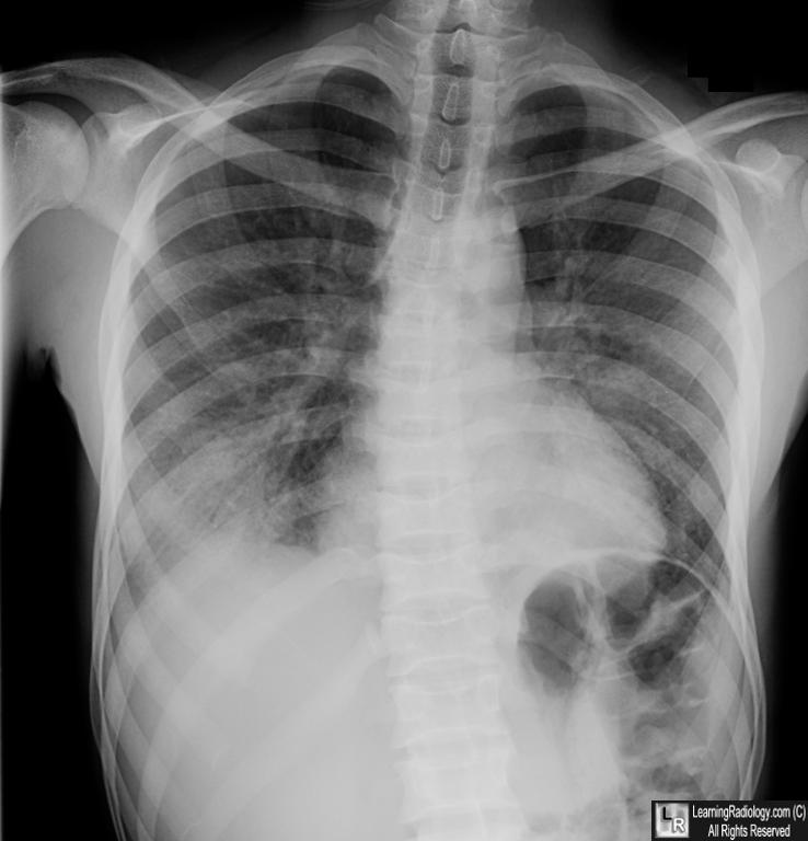
Acute Chest Syndrome. There is bilateral lower lobe airspace disease (white arrows). The patient has osseous changes of sickle cell disease including biconcave vertebral bodies (blue oval) and avascular necrosis of the visible humeral head (yellow arrow)
For this same photo with the arrows, click here
For more information, click on the link if you see this icon 
|
|
|
