Learning Radiology xray montage


Pneumomediastinum
Mediastinal Emphysema


  • Rare in adults (except in ICU patients) and most common in newborns

  • ETIOLOGIES

    • Spontaneous – most common mechanism in kids and adults

      • Pathophysiology involves increased intralveolar pressure such as from cough or vomiting which ruptures alveolus, air tracks back along blood vessels to mediastinum

        • Accompanying pneumothorax common in neonates

      • When air builds up in mediastinum and can’t pass into the neck (especially in kids) this produces mediastinal air block which can reduce flow of blood in great vessels

      • In adults, the air in mediastinum usually progresses into the neck and the subcutaneous tissue

·       Traumatic – 2° closed chest trauma                       

o      Same mechanism as spontaneous  

·       Rupture of the esophagus – Boerhaave's Syndrome                       

o      May occur with vomiting, labor, severe asthmatic attacks and strenuous exercise (each of these can produce pneumomediastinum without rupturing the esophagus)  

o      LEFT, POSTEROLATERAL WALL,  DISTAL 8 CM  

      • X-ray – combination of pneumomediastinum and left pleural effusion is very suggestive; continuous diaphragm sign

      • Symptoms – infants – none. 

      • Adults – chest pain (retrosternal) radiating down both arms   aggravated by respiration and swallowing; Hamman’s sign – crunching sound heard over the apex of the heart with cardiac cycle
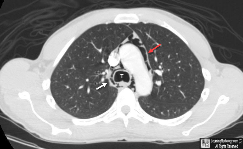
Pneumomediastinum

Pneumomediastinum. There is air in the mediastinum surrounding the aorta (red arrow) and trachea (T) (white arrow). The patient was an asthmatic who presumably ruptured a bleb with air dissecting back along the bronchovascular bundles of the lung to the mediastinum.