|
|
Calcification of the Ligamentum Arteriosum
General Considerations
- The ligamentum arteriosum is the remnant of the ductus arteriosis
- Calcification of the ligament may be seen a few months to several years after closure
- Relatively common finding on unenhanced CT of chest
- Prevalence increases with advancing age and atherosclerosis
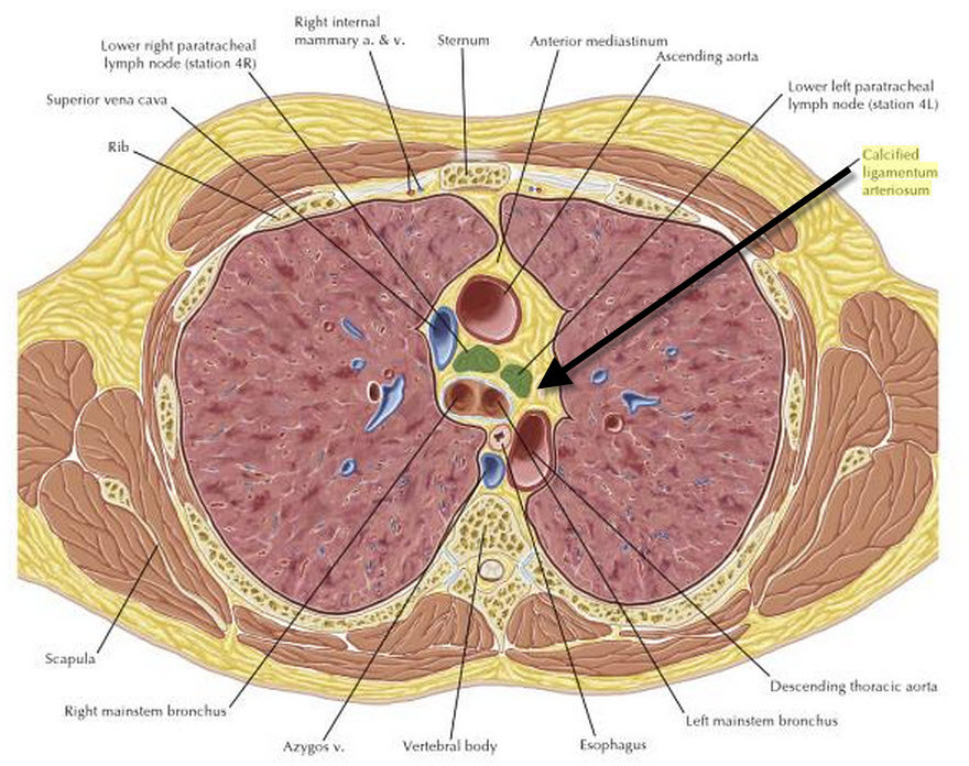
From Netter's Correlative Imaging by Gotway. Location of Ligamentum arteriosum.
Mouseover to enlarge.
Clinical Findings
- Calcification is almost always clinically insignificant
- It may be associated with long-standing patent ductus in adults
Imaging Findings
- While such calcification can be seen on conventional radiographs, the yield is low (3.6% in children)
- Calcification is more easily seen on spiral CT in the aorto-pulmonary window, connecting the top of the left pulmonary artery with the floor of the aortic arch
- Calcification can be punctate (most common), curvilinear or clumped
Differential Diagnosis
- Foreign body
- Granulomatous infection
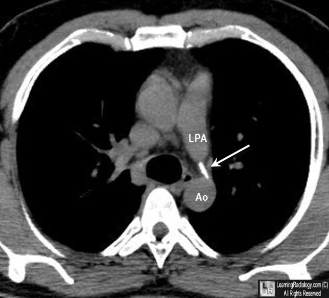
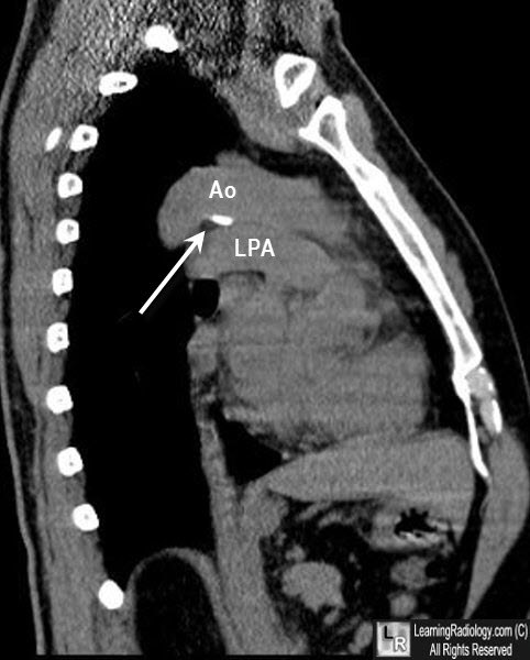
Calcification of the Ligamentum Arteriosum. Upper and lower photos of axial and sagittal unenhanced CT scans of the chest show calcified ligamentum (white arrow) connecting the left pulmonary artey (LPA) and aorta (Ao).
Calcification of the ligamentum arteriosum in adults: CT features. Wimpfheimer O1, Haramati LB, Haramati N. J Comput Assist Tomogr. 1996 Jan-Feb;20(1):34-7.
Prevalence of ligamentum arteriosum calcification on multi-section spiral CT and digital radiography. Gil-Sun Hong, Hyun Woo Goo, Jae-Woo Song. The International Journal of Cardiovascular Imaging, June 2012, Volume 28, Issue 1 Supplement, pp 61-67
|
|
|
