|
|
Tumoral Calcinosis
-
Progressively enlarging,
juxta-articular, calcified, nodular soft-tissue masses
-
Mostly occurs in 1st or 2nd decade
-
Equal M:F
-
Normal calcium and phosphorous
-
Autosomal dominant with variable
expressivity
-
Pathology: multilocular cystic lesions
containing creamy white fluid
-
Clinical
-
Imaging
-
Large, nodular, smoothly-marginated
juxta-articular masses of calcium density
-
Fluid-fluid levels on erect films due
to Milk of Calcium in lesion
-
Underlying bone normal
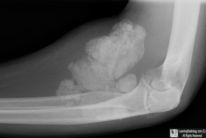
Tumoral Calcinosis. There is a well-circumscribed and multi-lobular area of increased density consistent with calcification in the soft tissue of the anterior forearm near the elbow joint. The density has an almost tumor-like appearance. The underlying bone is normal in appearance.
|
|
|
