|
|
Septic Arthritis
Infectious Arthritis
Infectious Arthritis
- Infectious Arthritis usually occurs as a result of hematogenous seeding of the synovial membrane from an infected source elsewhere in the body, like a wound infection or from direct, contiguous extension from osteomyelitis adjacent to the joint.
- It is usually subdivided into pyogenic (septic) arthritis, due mostly to Staphylococcal and Gonococcal organisms and non-pyogenic arthritis, due mostly to infection with Mycobacterium tuberculosis.
- Risk factors include intravenous drug use, steroid injections or oral administration, joint prostheses and recent joint trauma, including joint surgery.
- In children and adults, the knee is frequently affected; in children, the hip is another common site of infection. Hands may be infected from human bites and the feet from diabetes.
- Although conventional radiographs are obtained as the initial study, they are relatively insensitive to the early findings of the disease except for soft tissue swelling and osteopenia.
- If septic arthritis is strongly suspected, aspiration of the joint will usually confirm the diagnosis.
- The hallmark of infectious arthritis, especially the pyogenic form, is destruction of the articular cartilage and long, contiguous segments of the adjacent articular cortex from proteolytic enzymes released by the inflamed synovium.
- This destruction, unlike other arthridities, is usually quite rapid.
- Infectious arthritis tends to be monarticular and is associated with soft tissue swelling and osteopenia from the hyperemia of inflammation
- Because of its sensitivity, MRI is now used extensively after conventional radiography, in diagnosing septic joints. Enhancement of the synovium and the presence of a joint effusion have the best correlation with the clinical diagnosis of a septic joint.
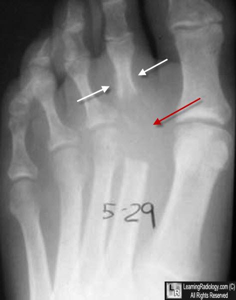
Septic Arthritis, 2nd toe. There is complete destruction of the 2nd metatarsophalangeal joint space (red arrow) with periosteal new bone formation along the shaft of the proximal phalanx (white arrow).
|
|
|
