|
|
Schmorl's Nodes
Cartilaginous Nodes
General Considerations
- Common (1/3 of population); more common in men than women
- Extrusion, invagination, protrusion of intervertebral disc material through a break in the subchondral end plate of a vertebral body
- The disk material is displaced into the vertebral body
- Most common in thoracolumbar spine
- May occur spontaneously or in association with
- Scheuermann’s disease (juvenile kyphosis)
- Trauma
- Hyperparathyroidism
- Osteoporosis, other metabolic conditions
- Infection
- Neoplasm
Clinical Findings
- Are generally considered to be asymptomatic although cases are reported of acute pain
Imaging Findings
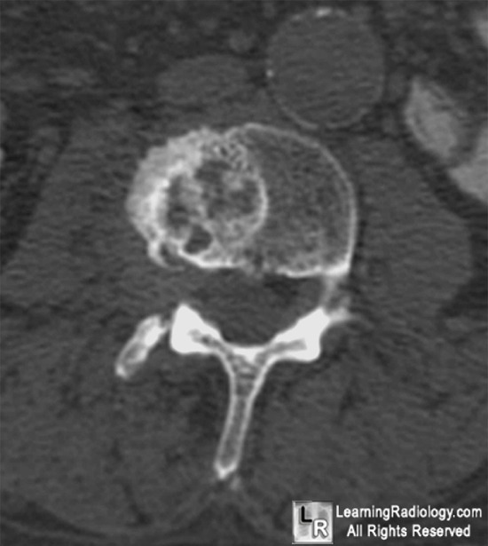
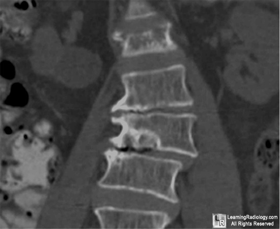
- Lucent lesion, most often in middle or posterior portion of vertebral body
- At margin with disc space
- More commonly involves inferior endplate
- Usually posterior of middle portion of endplate
- Sclerotic margin
- On MR
- Schmorl’s node generally has same signal characteristics as adjacent disk
- Best seen on sagittal images
- Defect in endplate is often seen
Differential Diagnosis
Treatment
- Usually asymptomatic
- If symptomatic, conservative symptomatic treatment


Schmorl's node. Black arrow points to well defined marginal lesion in L3 with sclerotic margins. There is associated degenerative osteophyte formation (white arrow). On coronal reconstruction, the marginal lesion is seen (blue arrow) and there is a vacuum disk phenomenon present from the degenerated disk below.
For more information, click on the link if you see this icon 
For this same photo without the annotations, click here and here
|
|
|
