|
|
Carpal Instabilities
|
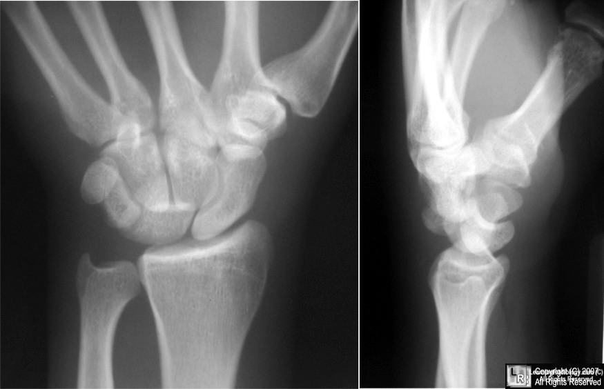
Lunate Dislocation
-
Most severe of
carpal instabilities
-
Most commonly
associated with a trans-scaphoid fracture
-
Involves all the
intercarpal joints and disruption of most of the major
carpal ligaments
-
Produces volar
dislocation and forward rotation of lunate
- Concave distal
surface of lunate comes to face anteriorly
-
Capitate drops
into space vacated by lunate
-
Capitate and all
other carpal bones lie posterior to lunate on lateral
radiograph
-
Triangular
appearance of lunate on frontal projection
|
|
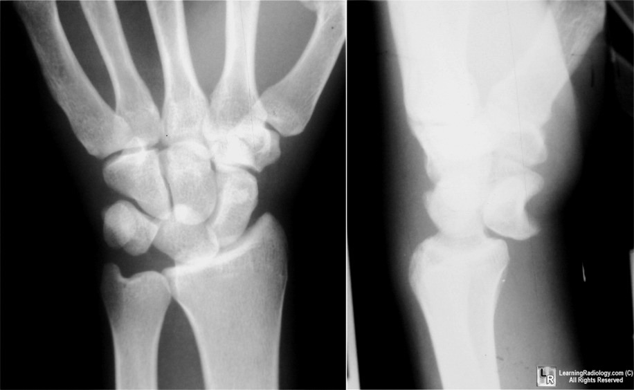
Perilunate Dislocation
- Result of a fall
on an outstretched, hyperextended hand
- Relatively rare
- Occurs when the
lunate maintains normal position with respect to the distal
radius while all other carpal bones are dislocated
posteriorly
-
Very commonly
associated with a scaphoid waist fracture
- Sometimes
ulnar styloid as well
- Lunate appears
triangular in shape on frontal view
- Lunate rotates
forward slightly on lateral view
- In lateral view,
all other carpal bones are dislocated posterior with respect
to lunate
|
|
Lunate and Perilunate Dislocation
- Carpal dislocations described by extent of
ligamentous injury (Mayfield)
- Stage I
- Isolated rotatory subluxation of scaphoid
- Mechanism: acute dorsiflexion of wrist
- May be associated with rheumatoid
arthritis
- Characterized by increased distance
between scaphoid and lunate > than 2 mm (Terry Thomas sign)
- Scaphoid ring sign – ring-shaped shadow of
cortex of distal pole of scaphoid seen on end
- Associated more than 50% of the time with
distal radial fractures
- Stage II
- Dislocation or subluxation of capitate
- Stage III
- Perilunate dislocation
- Triquetral and scaphoid malrotation
- Result of a fall on an outstretched,
hyperextended hand
- Relatively rare
- Occurs when the lunate maintains normal
position with respect to the distal radius while all other
carpal bones are dislocated posteriorly
- Very commonly associated with a scaphoid
waist fracture
- Sometimes ulnar styloid as well
- Lunate appears triangular in shape on
frontal view
- Lunate rotates forward slightly on lateral
view
- In lateral view, all other carpal bones
are dislocated posterior with respect to lunate
- Stage IV
- Lunate Dislocation
- Most severe of carpal instabilities
- Most commonly associated with a
trans-scaphoid fracture
- Involves all the intercarpal joints and
disruption of most of the major carpal ligaments
- Produces volar dislocation and forward
rotation of lunate
- Concave distal surface of lunate comes
to face anteriorly
- Capitate drops into space vacated by
lunate
- Capitate and all other carpal bones lie
posterior to lunate on lateral radiograph
- Triangular appearance of lunate on frontal
projection
|
|
|