|
|
Paget Disease of the Spine
General Considerations
Sites of involvement
- Usually polyostotic and asymmetric
- Pelvis (75%) most common, followed by
- Lumbar spine
- Thoracic spine
- Proximal femur
- Calvarium
- Scapula
- Distal femur
- Proximal tibia
- Proximal humerus
Imaging Findings
- Classical triad
- Thickening of the cortex
- Accentuation of the trabecular pattern
- Increased size of bone
- Cyst-like areas
- Skull (involvement in 29-65%)
- Inner and outer table involved
- Leads to diploic widening
- Osteoporosis circumscripta is well-defined lysis, most commonly in frontal bone producing well-defined geographic lytic lesion in skull
- Represents early destructive phase of disease active stage)
- "Cotton wool" appearance represents mixed lytic and blastic pattern of thickened calvarium (later stage)
- Basilar invagination with encroachment on foramen magnum
- Deossification and sclerosis in maxilla
- Sclerosis of skull base
- Long bones (almost invariably starts at end of bone)
- "Candle flame" or "blade of grass" pattern of lysis is the advancing tip of V-shaped lytic defect in diaphysis of long bone originating in subarticular site
- Lateral curvature of femur
- Anterior curvature of tibia (commonly resulting in fracture)
- Pelvis
- Thickened trabeculae in sacrum, ilium
- Rarefaction in central portion of ilium (looks like a large lytic lesion)
- Thickening of iliopectineal line
- Acetabular protrusio with secondary degenerative joint disease
- Spine (upper cervical, low dorsal, midlumbar most common sites)
- Coarse trabeculations at periphery of bone
- "Picture-frame vertebra" mimics bone-within-bone appearance
- Enlarged vertebral body with reinforced peripheral trabeculae and more lucent center, typically in lumbar spine
- "Ivory vertebra" is a blastic vertebra with increased density
- Ossification of spinal ligaments, paravertebral soft tissue, disk spaces can occur
MRI Findings
- Hypointense area / area of signal void on T1WI + T2WI (cortical thickening, coarse trabeculation)
- Widening of bone
- Reduction in size and signal intensity of medullary cavity due to replacement of high-signal-intensity fatty marrow by medullary bone formation
- Focal areas of higher signal intensity than fatty marrow (from cyst-like fat-filled marrow spaces)
- Areas of decreased signal intensity within marrow on T1WI and increased intensity on T2WI (= fibrovascular tissue resembling granulation tissue)
DDx
- Depends on the bone in which it occurs
- Skull
- Osteolytic or osteoblastic metastases
- Long bones
- Metastases
- Chronic osteomyelitis (thickened cortex)
- Old trauma (thickened cortex)
- Hodgkin’s disease
- Spine
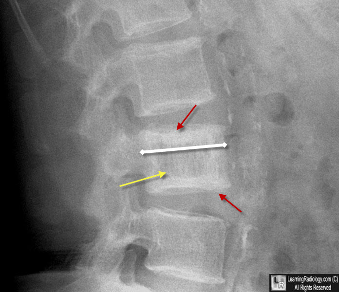
Paget Disease of Spine. The lumbar vertebral body has thickened cortices which outline the body ("picture-frame appearance") (red arrows). The vertebral body is slightly larger than the body above and below it (white double arrow). The trabecular pattern is thickened and coarsened (yellow arrow).
|
|
|
