|
|
Paget Disease of the Rib
General Considerations
- Multifocal chronic skeletal disease due to chronic paramyxoviral infection
- Prevalence
- 3% of individuals >40 years
- 10% of persons >80 years
- Unusual <40 years
- M:F = 2:1
- Pelvis most commonly involved
- Histology
- Increased resorption and increased bone formation
- Newly formed bone is abnormally soft with disorganized trabecular pattern
Clinical Findings
- Asymptomatic (1/5)
- When symptomatic, symptoms may include
- Fatigue
- Enlarged hat size
- Peripheral nerve compression
- Neurologic disorders from compression of brainstem (basilar invagination)
- Hearing loss, blindness
- Facial palsy (narrowing of neural foramina) - rare
- Pain from(a)primary disease process is rare so think of
- Pathologic fracture
- Malignant transformation
- Secondary degenerative joint disease aggravated by skeletal deformity
- High-output congestive heart failure from markedly increased perfusion (rare)
- Increased alkaline phosphatase (increased bone formation)
- Hydroxyproline increased (increased bone resorption)
- Normal serum calcium + phosphorus
Imaging Findings
- Characteristic changes of Paget’s disease of bone include
- Thickening of the cortex
- Coarsening of the trabecular pattern, and
- Increased size of the bone
Differential Diagnosis
- Metastatic disease
- Fibrous dysplasia
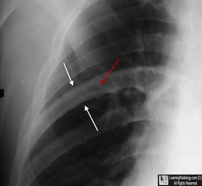
Paget Disease, right 6th rib. The rib is enlarged. The cortex is thickened (white arrows) but the medullary space remains intact (red arrow) with no evidence of infiltration.
|
|
|