|
|
Multiple Hereditary Exostoses
Diaphyseal Aclasis
- Inheritance
- Age of onset
- Discovered
between 2 and 10 years
- Male
predominance = 2:1
- Pathology
- Ectopic
cartilaginous rest in metaphysis and a defect in periosteum produces
exostoses
- Cap of
hyaline cartilage over bony protuberance
- Cortex and cancellous bone of exostosis is contiguous to host bone
- Clinical
- Usually
painless mass near joints
- Tendons,
blood vessels, nerves may be impaired
- Mechanical
limitation of joint movement may occur
- Location
- Multiple
- Usually
bilateral
- Common sites
are knee, elbow, scapula, pelvis, ribs
- Site
- Metaphyses
of long bones near epiphyseal plate (distance to epiphyseal line
increases with growth)
- Always point
away from joint and toward center of shaft
- Occasionally
small punctate calcifications are seen in cartilaginous cap
- Other
skeletal abnormalities may occur
- Shortening
of 4th and 5th metacarpals
- Supernumerary
fingers and/or toes
- Madelung / reversed Madelung deformity
- Dislocation
of radial head
- Prognosis
- Exostosis begins in childhood
- Stops
growing when nearest epiphyseal center fuses
- Complications
- Malignant
transformation to chondrosarcoma in <5%
- Iliac bone
commonest site
- Look for growth
with irregularity of contour and fuzziness of margin
- Sudden
painful growth spurt
- Cord
compression secondary to involvement of posterior spinal elements
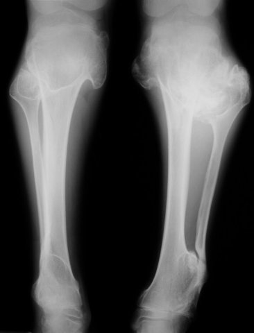
Multiple Hereditary Exostoses. Multiple exostoses are seen arising from the proximal and distal tibias and fibulas. The bones are dysplastic in appearance.
|
|
|
