|
|
Lisfranc Fracture Dislocation
- Named after Jacques Lisfranc, a field surgeon in Napoleon’s army, who described a new technique for an amputation used to treat frostbite of the forefoot in soldiers on the Russian front
- Used today to describe fractures and dislocations that occur at the junction between the tarsal bones of the midfoot and the metatarsals of the forefoot
- Causes
- Mechanism involves severe plantar flexion of the foot
- May occur from sports-related injuries
- Motor vehicle accidents
- Falling from a height, down stairs or off a curb
- Ligamentous injuries alone, even without fracture or dislocation, may result in instability on weightbearing
- Lisfranc ligament diagonally connects the 1st (medial) cuneiform with the base of the 2nd metatarsal
- If it remains intact, either an avulsion of the lateral border of the 1st cuneiform or an avulsion of the base or medial border of the 2nd metatarsal occurs
- If it tears, these fractures may not occur
- Clinical findings
- Pain at tarsal-metatarsal joints
- Ecchymosis
- Instability
- Types
- Two basic types
- Homolateral
- All of the metatarsals are dislocated to the same side
- More common than divergent
- Usually involves the 2nd through 5th dislocated laterally
- May involve all 5 metatarsals
- Divergent
- Usually more severe than homolateral
- May be associated with a fracture of the 1st cuneiform
- Usually involves medial displacement of the 1st metatarsal and lateral displacement of 2nd-5th metatarsals
- Occasionally may involve only medial displacement of only the 1st metatarsal
- Fractures associated with Lisfranc dislocations
- Base of 2nd metatarsal
- Cuboid
- Fractures of shafts of metatarsals
- Dislocations of the 1st (medial) and 2nd (middle) and cuneonavicular joints
- Fractures of the tarsal navicular
- Imaging
- Conventional radiographs are usually sufficient to demonstrate the injury
- Normal alignment of the cuneiforms and the bases of the metatarsals (see chart below)
- Lateral border of 1st metatarsal is aligned with lateral border of 1st (medial) cuneiform on AP view
- Medial border of 2nd metatarsal is aligned with medial border of 2nd (intermediate or middle) cuneiform on AP view
- Medial and lateral borders of the 3rd (lateral) cuneiform should align with medial and lateral borders of 3rd metatarsal on oblique view
- Medial border of 4th metatarsal aligned with medial border of cuboid on oblique
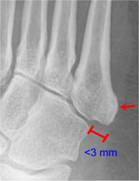
- Lateral margin of the 5th metatarsal may project lateral to cuboid by as many as 3mm on oblique
- On lateral, a line drawn along long axis of talus should intersect long axis of 5th metatarsal
Normal Alignment of Tarsal-Metatarsal Joints |
Metatarsal |
AP Projection |
Oblique Projection |
1st |
|
Lateral border of 1st metatarsal is aligned with lateral border of 1st (medial) cuneiform
|
2nd |
|
Medial border of 2nd metatarsal is aligned with medial border of 2nd (intermediate) cuneiform
|
3rd |
Medial and lateral borders of the 3rd (lateral) cuneiform should align with medial and lateral borders of 3rd metatarsal
|
|
4th |
Medial border of 4th metatarsal aligned with medial border of cuboid
|
|
5th |
Lateral margin of the 5th metatarsal can project lateral to cuboid by up to 3mm on oblique
|
|
On lateral view |
Line drawn along long axis of talus should intersect long axis of 5th metatarsal |
- Stress views of the foot with the patient sedated will usually demonstrate any instability
- Lisfranc dislocations may be missed in up to 20% of cases
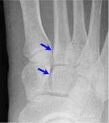
- Suspect it is present if there is a gap of more than 5 mm between bases of 1st and 2nd metatarsals or 1st (medial) and 2nd (middle or intermediate) cuneiforms
- On lateral view, bones of the midfoot will be subluxed or dislocated in a plantar direction
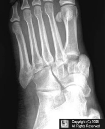
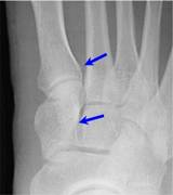
Lisfranc Fracture-Dislocation. The bases of all of the metatarsals
are dislocated laterally in this homolateral Lisfranc dislocation. There was
a fracture of the base of the 2nd metatarsal.
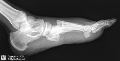
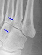
Lateral view of Lisfranc dislocation. Notice how the bones of the midfoot are dislocated
towards the plantar aspect of the foot.
|
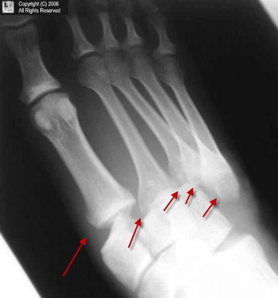
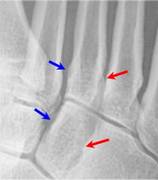
Lisfranc Dislocation. There is lateral dislocation of all of the metatarsals on their corresponding tarsal bones (red arrows).
- Treatment
- Sprains with an otherwise stable tarsal-metatarsal joint can be managed with immobilization
- Nonanatomic alignment requires open reduction and internal fixation
|
|
|
