|
|
Erosive Osteoarthritis
Inflammatory Osteoarthritis
General Considerations
- Inflammatory arthritis which most commonly occurs in women over the age of 60
- It is a form of osteoarthritis with a strong inflammatory component
- Usually involves hand
- Hip and knee are rarely involved
Clinical Findings
- Onset is more acute than typical osteoarthritis
- Swelling
- Erythema
- Warmth
- Tenderness
- Usually begins in DIP joints and progresses to PIP joints
- Typically rheumatoid factor negative
Imaging Findings
- Typically bilateral, poly-articular and relatively symmetrical involvement
- Central erosions are a hallmark resulting in gull-wing appearance
- Central erosion proximally with marginal proliferation distally at both the DIP and PIP joints
- Gull-wing configuration is not specific for erosive osteoarthritis and may be seen with rheumatoid and psoriatic arthritis
- Most frequently seen in DIP joints of hands
- Spares MCP joints
- Frequent involvement of the carpal-metacarpal joint of the thumb
- May produce cortical thickening similar to psoriatic arthritis
- Ankylosis may occur
- In the foot, erosive osteoarthritis may involve either the metatarsal-phalangeal joint or interphalangeal joint of the great toe
Differential Diagnosis
- Osteoarthritis – erosions and ankylosis usually not seen
- Rheumatoid arthritis – erosions are usually marginal
- Psoriatic arthritis – systemic findings, foot may be involved
Treatment
- Conservative treatment
- Course of the disease is not dramatically changed by any mode of therapy
Prognosis
- Good
- Inflammatory component frequently self-extinguishes leaving changes of typical osteoarthritis
- Has been associated with hypothyroidism, autoimmune thyroiditis, hyperparathyroidism, chronic renal disease, scleroderma, Sjögren's syndrome, and calcium pyrophosphate dihydrate arthropathy


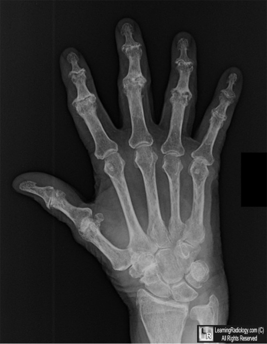
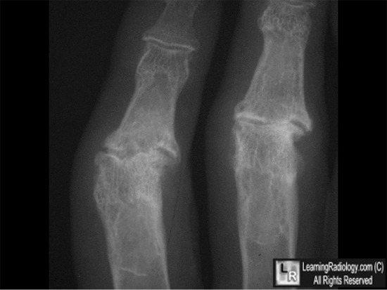
Erosive Osteoarthritis. (Above). Frontal radiograph of the hand demonstrates an arthritis which affects mainly the DIP and PIP joints (white arrows) and carpal-metacarpal joint of thumb (yellow arrow). There are small osteophytes and erosions (white circle). (Below) The characteristic lesion of erosive osteoarthritis is shown in close-up. There is a central erosion of the proximal part of joint (yellow arrow) and bone overgrowth peripherally (white arrows) resembling a seagull's wings.
For these same photos without the arrows, click here and here
For more information, click on the link if you see this icon 
Advanced Erosive Osteoarthritis. RS Daniel and AN Brown. Applied Radiology. 2007;36(12) © 2007
Erosive Osteoarthritis and Psoriatic Arthritis: A Radiologic Comparison in the Hand, Wrist, and Foot. W Mantel, KJ Stuck, A. Dwonin and RG HylIand. AJR 134:125-135, January 1980.
Erosive Osteoarthritis. KL Kidd and J B Peter. April 1966 Radiology, 86, 640-647.
|
|
|
