|
|
Enchondroma
- General considerations
- Benign cartilaginous tumor
- Develops in the medullary cavity
- Usually solitary, although they can occur as multiple lesions in syndromes to be discussed
- Arise from ectopic rests of hyaline cartilage
- Occur mostly in 2nd to 3rd decade
- Most common site is small bones of the hands and feet
- Most occur in the proximal phalanx
- Most common tumor of the hand
- Also occur in the humerus, femur, tibia and ribs
- In long bones, their calcification is similar to an intramedullary bone infarct
- Bone infarcts tend to have a well-circumscribed, sclerotic margin
- May fracture or, rarely, undergo malignant transformation
- On CT, lucent regions in an otherwise dense enchondroma suggest malignancy
- Rapid growth of lesion or pain not related to a pathologic fracture may suggest malignant transformation
- Clinical findings
- Usually asymptomatic and found serendipitously
- May be associated with pain and swelling which should raise suspicion of either a pathologic fracture or, less likely, malignant transformation
- Malignant transformation almost never occurs in the hands and feet
- Imaging findings
- Conventional radiography is the study of first choice
- Well-defined lytic and slightly expansile lesion (in small bones)
- Usually have some internal calcification and endosteal thinning
- Internal calcifications tend to resemble “rings and arcs” of cartilage calcification
- MRI findings
- Numerous internal foci with high-signal intensity on T2
- Low to intermediate signal on T1
- Lobulated in contour
- Bone scan
- Negative if uncomplicated enchondroma
- Multiple enchondromas occur in Ollier's disease
- Nonhereditary
- Frequently unilateral
- Enchondromas are frequently larger than with solitary enchondroma
- May have limb shortening of affected limb
- May have Madelung’s deformity of the wrist
- Greater incidence of malignant transformation because there are more lesions present (25-50%)
- Enchondromas associated with cavernous hemangiomas of soft tissues is called Maffucci’s Syndrome
- Nonhereditary
- Even more rare than Ollier’s disease
- Multiple hemangiomas usually in extremities (digits)
- Look for phleboliths in hemangioma
- Growth disturbance of affected bones
- Malignant transformation
- Greater than solitary enchondroma, less than Ollier’s
- Hemangioma may become sarcoma in 5% of cases
 
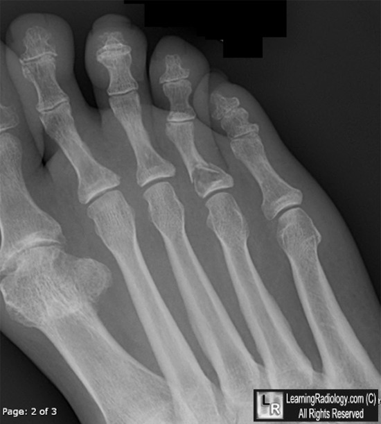
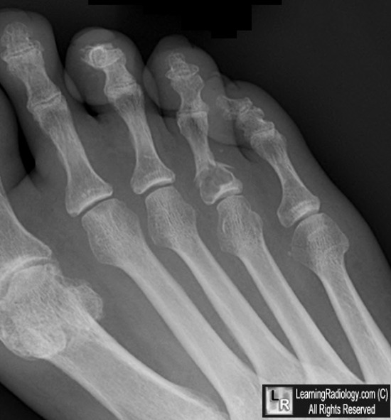
Enchondroma with pathologic fracture. Left: There is a non-displaced fracture through the base
of the proximal phalanx of the 4th toe (white arrow). Right: A well-defined, slightly expansile, lytic lesion
is seen in the proximal phalanx (white circle) through which the fracture has occurred.
For these same photos without the arrows, click here and here
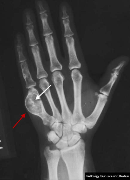
Enchondroma. There is an expansile, lytic lesion of the 5th metacarpal (red arrow) containing amorphous matrix calcifications (white arrow) characteristic of an enchondroma. There is no pathologic fracture.
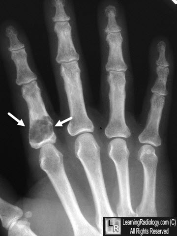
Enchondroma. There is an expansile, lytic lesion of the metacarpal of the index finger (white arrows) containing amorphous matrix calcifications.
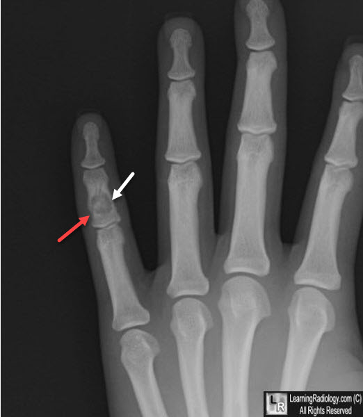
Enchondroma. There is a lytic lesion of the middle phalanx of the ring finger (white arrow) with amorphous calcified matrix within it. A pathological fracture is also present (red arrow).
For more information, click on the link if you see this icon 
|
|
|
