|
|
Calcinosis of Chronic Renal Disease
Secondary Tumoral Calcinosis
General Considerations
- Also called uremic tumoral calcinosis and sometimes secondary tumoral calcinosis
- Calcified masses are uncommon overall with a frequency range between 0.5% and 3% in renal patients, but the most frequent cause of a calcified periarticular “mass” is chronic renal failure
- Occurs more commonly in patients on hemodialysis for greater than 3 years
- No histologic or radiologic differences between this type of calcinosis and the lesions of primary tumoral calcinosis
Clinical Findings
- Most frequently occurs in the context of hyperparathyroidism
Imaging Findings
- Amorphous and/or multi-lobulated calcification located in a periarticular distribution
- Joint space is preserved
- Underlying bone and muscle are not usually involved
- CT shows no erosion or adjacent destruction of bone
- MRI shows inhomogeneous high-signal intensity on T2-weighted sequences while T1-weighted sequences usually show inhomogeneous lesions with low signal intensity
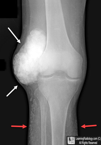
Calcinosis of Chronic Renal Failure. A smoothly marginated, very dense and multilobulated calcified mass is seen adjacent to the right knee joint (white arrows). There is also diffuse soft tissue calcification present (red arrows.
Tumoral Calcinosis: Pearls, Polemics and Alternative Possibilities. KM Olsen and FS Chew. RadioGraphics. May-June, 2006, Vol. 26, 3.
|
|
|
