|
|
Intraosseous Lipoma of the Calcaneus
General Considerations
- Uncommon, and not as rare as once thought
- Benign tumor composed of mature, adult fat
- Adults between 30-60
- Males slightly more than females
- Frequent skeletal locations include
- Proximal femur, intertrochanteric region (34%)
- Base of neck of calcaneus (8%)
- Tibia (13%)
- Ilium (8%)
Clinical Findings
- Found incidentally 1/3 time
- Pain
- Tenderness
- Swelling
Milgram's Classification Intraosseous Lipomas |
Stage |
Description |
1 |
Viable fat without necrosis |
2 |
Viable fat with necrosis and dystrophic calcification |
3 |
Extensive fat necrosis, calcification, cysts |
Imaging Findings
- On MRI
- Fat in lipoma is isointense with subcutaneous fat on T1
- Low signal on T1 with fat suppression
- On conventional radiographs
- Lucent lesion with sclerotic margin
- Central sclerotic bulls-eye (pathognomonic)
- Cockade image-resembles the badge or rose worn upon a hat
- May be associated with mild expansion of long bone
Differential Diagnosis
- Not to be mistaken for normal areas of lucency in calcaneous and proximal femur
- Osteonecrosis
Treatment
- None usually needed if asymptomatic
- Curettage and bone grafting for symptomatic lesions
Complications
- Recurrence and malignant transformation is very rare
Prognosis
- Frequently disappears spontaneously
 
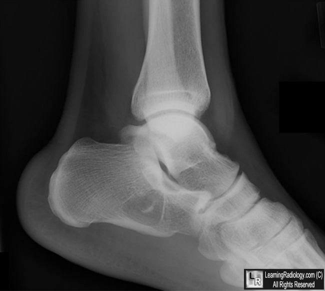
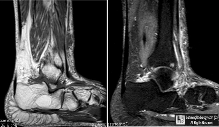
Intraosseous Lipoma of the Calcaneus. Lateral conventional radiograph demonstrates a classical
well-defined lytic lesion in the calcaneus with a sclerotic margin (black arrow) and a central calcification (white arrow). Sagittal T1 and T1 STIR images demonstrate a fat-containing lesion (black and white arrows).
For more information, click on the link if you see this icon 
For these same photos without the annotations, click here and here
Intraosseous lipoma of the calcaneus. Christoph Bertram, Frank Popken and Jürgen Rütt
Archives of Surgery: Volume 386, Number 5 / August, 2001
Benign Musculoskeletal Lipomatous Lesions. Mark D. Murphey, MD, John F. Carroll, MD, Donald J. Flemming, CAPT, MC, USN, Thomas L. Pope, MD, Francis H. Gannon, MD and Mark J. Kransdorf, MD. September 2004 RadioGraphics, 24, 1433-1466.
|
|
|
