|
|
Olecranon Bursitis - Gout
General Considerations
- Deposition of sodium urate monohydrate crystals in synovial membranes, articular cartilage, ligaments, bursae leading to destruction of cartilage
- Age of onset is usually greater than 40 years; males much more often than females
- Radiologic features usually not seen until 6-12 years after initial attack
- Radiologic features present in 50% of affected patients
Causes:
- M:F = 20:1
- Overproduction of uric acid
- Abnormality of renal urate excretion
- Rarely cause for radiographically apparent disease
- Myeloproliferative disorders, e.g. polycythemia vera, leukemia, lymphoma, multiple myeloma
- Blood dyscrasias
- Myxedema, hyperparathyroidism
- Chronic renal failure
- Glycogen storage disease
- Myocardial infarction
- Lead poisoning
Stages:
- Asymptomatic hyperuricemia
- Acute mono-articular gout
- Polyarticular gout
- Chronic tophaceous gout = multiple large urate deposits
Location:
- Joints: hands + feet (1st MTP joint most commonly affected = podagra), elbow, wrist
- Carpometacarpal compartment especially common), knee, shoulder, hip, sacroiliac joint (15%, unilateral)
- Ear pinna > bones, tendon, bursa
Soft tissue findings
- Calcific deposits in gouty tophi in 50% (only calcium urate crystals are opaque)
- Eccentric juxta-articular lobulated soft-tissue masses (hand, foot, ankle, elbow, knee)
- Bilateral olecranon bursitis
- Aural calcification
Joint findings
- Preservation of joint space initially
- Absence of periarticular demineralization
- Erosion of joint margins with sclerosis
- Cartilage destruction late in course of disease
- Periarticular swelling (in acute mono-articular gout)
- Chondrocalcinosis (menisci, articular cartilage of knee) resulting in secondary osteoarthritis
Bone findings
- "Punched-out" lytic bone lesion ± sclerosis of margin
- "Mouse / rat bite" from erosion of long-standing soft-tissue tophus
- "Overhanging margin" (40%)
- Ischemic necrosis of femoral / humeral heads
- Bone infarction
Coexisting disorders:
- Psoriasis
- Glycogen storage disease Type I
- Hypo- and hyperparathyroidism
- Down’s syndrome
- Lesch-Nyhan syndrome (choreoathetosis, spasticity, mental retardation, self-mutilation of lips + fingertips)
Treatment
- Colchicine, allopurinol (effective treatment usually does not change x-ray findings)

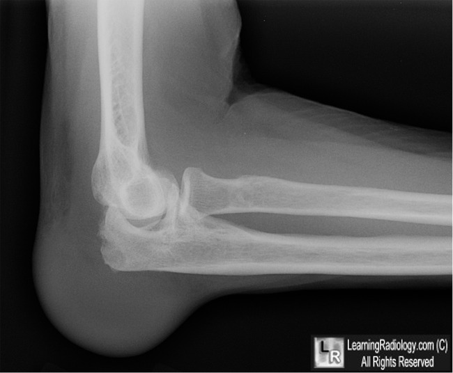
Olecranon Bursitis-Gout. The olecranon bursa is enlarged white arrow) and there is a large erosion in the
olecranon process of the ulna (yellow arrow).
For this same photo without the arrows, click here
For more information, click on the link if you see this icon 
|
|
|
