|
|
Avascular Necrosis of the Humeral Head
General Considerations
- Second most common joint for avascular necrosis (AVN) to hip
- Causes include
- Mechanical disruption from trauma
- Steroid-induced disease
- Sickle cell disease
- Alcohol abuse
Clinical Findings
- Insidious onset of pain
- Pain, poorly localized and usually severe
- Night and rest pain
- Range of motion is initially preserved
Cruess Classification |
Stage |
Findings |
Stage I |
Normal x-ray. Changes on MRI |
Stage II |
Sclerosis (wedged, mottled), osteopenia |
Stage III |
Crescent sign indicating a subchondral fracture |
Stage IV |
Flattening and collapse |
Imaging Findings
- Radiographs will be normal early in disease
- Then resorption in superior middle portion of humeral head
- Crescent sign (lucency) consistent with subchondral collapse
- Relative increase in density of head
- Can progress to osteoarthritic changes of joint eventually
- MRI preferred imaging modality
- Subchondral edema
- Low signal serpiginous line
- Double line sign (inner bright line from granulation tissue and outer dark line from sclerotic bone) on T2-weighted images
Differential Diagnosis
- Osteoarthritis involves both sides of the joint
Treatment
- Conservative treatment includes pain medication, physical therapy
- Operative procedures from core decompression to total shoulder arthroplasty
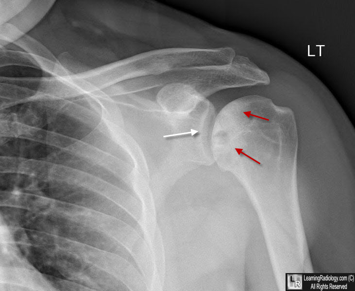
Avascular Necrosis of the Humeral head. There is a relative increase in density in the humeral head (white arrows) with a subchondral lucency seen in the medial portion of the head. The shoulder joint space is still preserved (red arrow).
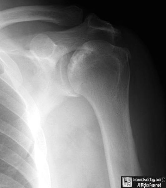
Avascular Necrosis of the Humeral head. There is a relative increase in density in the humeral head with irregularity in the surface of the head. The shoulder joint space is still preserved.
|
|
|
