
RecognizingA Pneumothorax
© William Herring, MD, FACR
Remember
There are two layers of pleura- parietal andvisceral-the pleural space between them
Normally there is no air in the pleuralspace
The visceral pleura is inseparable from thelung parenchyma and moves with the lung
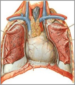
Visceralpleura
Parietalpleura
Pleuralspace
© Frank Netter, MD Novartis®
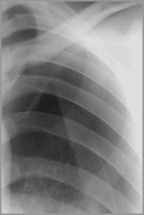
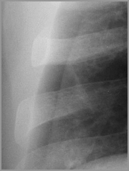
The Visceral Pleural White Line
When air enters the pleural space, theparietal and visceral pleura separatemaking the visceral pleura visible
The thin white line of the visceral pleurais called the visceral pleural white line
You must see the visceral pleural whiteline to make diagnosis of pneumothorax!
A pneumothoraxwill be visible as athin white line - thevisceral pleuralwhite line
Lung Markings
Lung markings may be absent distal to thevisceral pleural white line
But they can be seen distal to the visceralpleural white line even with apneumothorax if lung is folded on itself
Absence of lung markings is not sufficientto make diagnosis of pneumothorax!
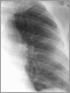
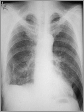
Large bulla in the LLL. Note there is novisceral pleural white line paralleling the chest wall
ARRS R3 ©
Why The Pleural White LineIs Important
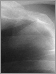
Chest tube erroneously inserted into bullain LUL produces an intractable pneumothorax.
ARRS R3 ©
Why The Pleural White LineIs Important
There are diseases other than apneumothorax that can cause anabsence of lung markings
For example
Bullous disease
Large cysts in the lung
Pulmonary embolism
Why The Pleural White LineIs Important
Why The Pleural White LineIs Important
None of those diseases is treated with achest tube
In fact, insertion of a chest tube into abulla can produce an intractablepneumothorax
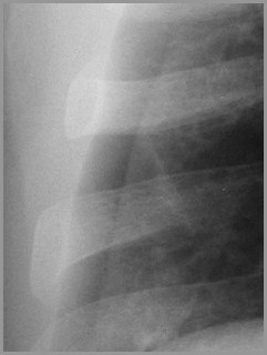
Skin fold or Pneumothorax
A fold of the patient’s skin may becometrapped between the patient and cassette
Skin folds are common
Especially in patient’s who have lost a greatdeal of weight
This skin fold can mimic a pneumothorax
How can we tell them apart?
Skin FoldSkin Fold
Pneumothorax
The key difference is that a skin fold is an edgeconsisting of a density (light) and then a lucency (dark)
Skin Fold
Dense
Lucent
This is an edge
Whereas the visceral pleural line is athin white line with a lucency (darker) on both sides of it
Pneumothorax
Lucent
Dense
Lucent
This is a line
Here they are again side-by-side: the skin fold is an edge, the pneumothorax is a line
Skin FoldSkin Fold
Pneumothorax
Which is this?Skin fold or Pneumothorax
This is anedge =skin fold
Which is this?Skin fold or Pneumothorax
This is a line =pneumothorax
Types of Pneumothoraces
Two major types of pneumothorax
Simple
Tension
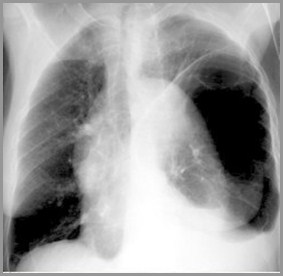
Simple Pneumothorax
In a simple pneumothorax, there is noshift of the heart or mediastinalstructures (trachea)
Air in left hemithorax balances the air inthe right hemithorax
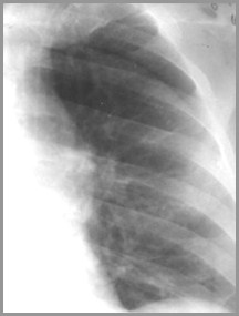
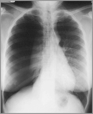
Simple pneumothorax on the left sideNo shift of the heart or trachea
Visceralpleural whiteline
No shift ofmidlinestructures
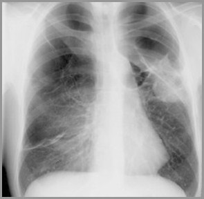
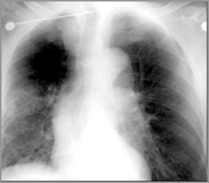
Tension Pneumothorax
Progressive loss of air into pleural spacecausing a shift of the heart and mediastinalstructures away from side of pneumothorax
Opposite lung is compressed
Respiratory function severely compromised
Tension Pneumothorax
© Frank Netter, MD Novartis®
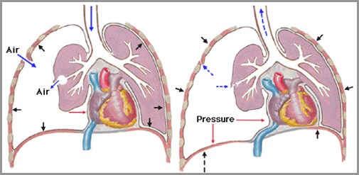
Air enters Right hemithorax either from tear in lung or hole in chestwall on inspiration; does not exit on expiration
Complete right-sidedpneumothorax
Lung iscompressedagainstmediastinum
Shift ofheart andtrachea toleft
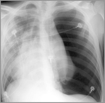
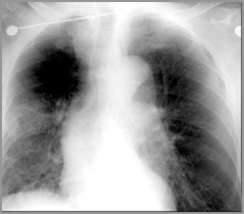
Which is this?Simple or Tension Pneumothorax
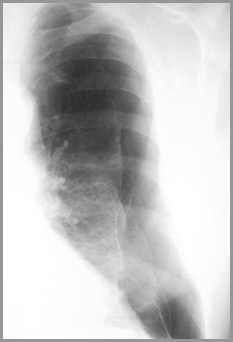
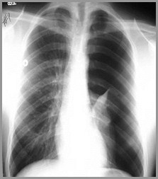
Tension pneumothorax-heart is shifted slightlyto right by large left-sided pneumothorax
Shifts with a Pneumothorax
If simple, there is no shift of heart and/ortrachea
If tension, there is a shift of heart and/ortrachea away from side of pneumothorax
There is never a shift toward the side of apneumothorax
Causes of a Pneumothorax
Spontaneous
Rupture of an apical sub-pleural bleb usuallyin a tall, thin male
Trauma
Through chest wall, e.g. stab wound
Internal, e.g. rupture of a bronchus from amotor vehicle collision
Causes of a Pneumothorax
Diseases that decrease lung compliance
Chronic fibrotic diseases, e.g. eosinophilicgranuloma
Diseases that stiffen the lung, e.g. hyalinemembrane disease
Rupture of an alveolus or bronchiole
E.g., asthma
Take Home Points
You must see the visceral pleural whiteline to diagnose a pneumothorax
A skin fold is an edge; the visceralpleural line is a line
There is a never a shift toward the sideof a pneumothorax


Congratulations, You Graduate

You know yourpneumothoraces
To repeat slide show, click here
To repeat only quiz cases, click here