
GastrointestinalRadiology
Differential Diagnoses
Reference and photos:Clinical Imaging, An Atlas of Differential Diagnosis, 2nd edition Eisenberg, RL
Submitted by Susan Summerton, MD
Submitted by Susan Summerton, MD
How This Works
•Eachdifferentialstarts with aphoto of thefindings
How This Works
•With the nextadvance, a blueclue will appearin the lowerright-handcorner to give ageneral idea ofthe differentialbeing asked

How This Works
•The next slidegives the title ofthe differentialand the numberof items on thelist
How This Works
•The last slidein each setgives thedifferential
Start
Start

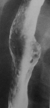
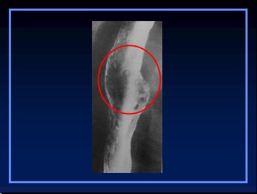
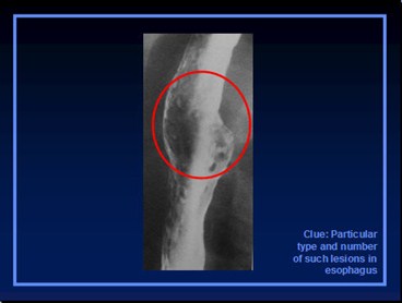
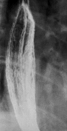
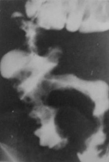
Clue: Particulartype and numberof such lesions inesophagus
Multiple Esophageal Ulcers
1.
2.
3.
4.
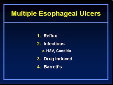
Multiple Esophageal Ulcers
1.Reflux
2.Infectious
a. HSV, Candida
3.Drug induced
4.Barrett’s
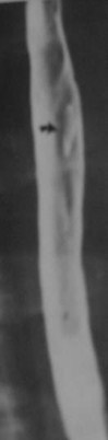
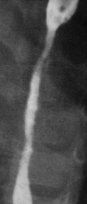
Clue: Particulartype and numberof such lesions inesophagus
Solitary Esophageal Ulcer
1.
2.
Solitary Esophageal Ulcer
1.HIV
2.CMV
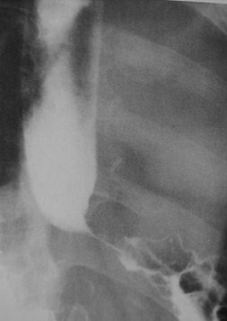
Clue: Particulartype of lesion andits location
Distal Esophageal Stricture
1.
2.
3.
4.
Distal Esophageal Stricture
1.Peptic strictures
2.Barrett’s
3.Carcinoma
a. Gastric,esophageal
4.Achalasia
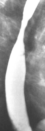
Clue: Particulartype of lesion andits location
Mid-esophageal stricture
1.
2.
3.
4.
Mid-esophageal stricture
1.Barrett’s
2.Radiation
3.Ingestion of corrosives
4.Metastases to mediastinum
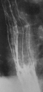
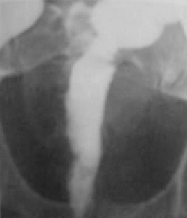
Clue: Particulartype of lesion
Esophageal Mucosal Nodularity
1.
2.
3.
4.
Esophageal Mucosal Nodularity
1.Reflux
2.Candida
3.Glycogenic Acanthosis
4.Barrett’s
Clue: Length ofthe lesion and itslocation
Long esophageal stricture
1.
2.
3.
Long esophageal stricture
1.Lye
2.NG Tube (prolonged use)
3.Radiation
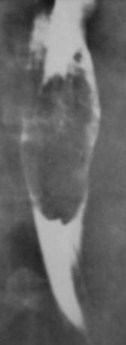
Clue: Nature andlocation of folds
Thickened esophageal folds
1.
2.
3.
4.
1.Varices
2.Varicoid carcinoma
3.Reflux esophagitis
4.Lymphoma
Thickened esophageal folds
Clue: Locationand number oflesions
Solitary esophageal mass
1.
2.
3.
4.
5.
Solitary esophageal mass
1.Leiomyoma
2.Fibrovascular polyp
3.Inflammatory esophagogastric polyp
4.Papilloma
5.Carcinoma
Clue: Type oflesion
Gastric ulcers, no mass
1.
2.
3.
4.
Gastric ulcers, no massCauses
1.H. Pylori
2.Aspirin
3.Crohn’s
4.Zollinger-Ellison syndrome
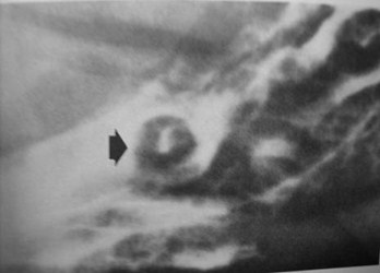
Clue: Type oflesion

Target lesions in GI tract
1.
2.
3.
Target lesions in GI tract
1.Hematogenous metastases
a. Breast, lung
2.Kaposi’s Sarcoma
3.Melanoma
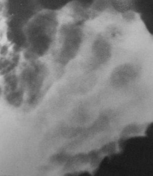
Clue: Nature oflesion in thisorgan
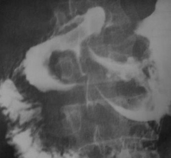
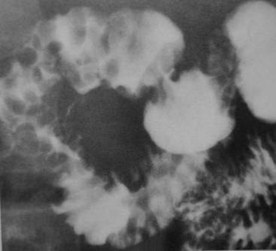
Thickened Gastric Folds
1.
2.
3.
4.
5.
1.Gastritis
a. Hypertrophic, H. Pylori
2.Menetrier’s disease
3.Zollinger Ellison syndrome
4.Varices
5.Lymphoma
Thickened Gastric Folds
Clue: Nature oflesion in thisorgan
Gastric Non-distensibility
1.
2.
3.
4.
Gastric Non-distensibility
1.Carcinoma
a. Primary, metastatic
2.Lymphoma
3.Atrophic gastritis
4.Scarring from PUD
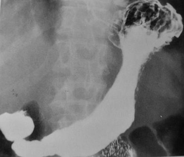
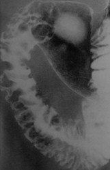
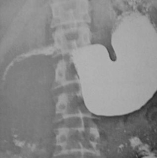
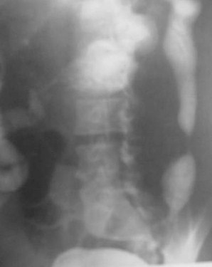
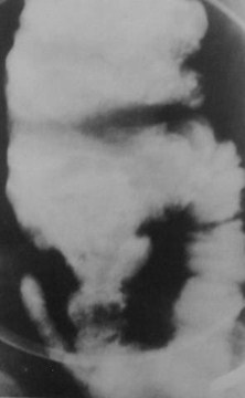
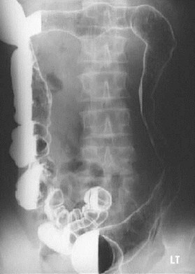
Linitis Plastica of the StomachDifferential Diagnosis
1.
2.
3.
4.
5.
6.
7.
Linitis Plastica
1.Pancreatic tumor
2.Lymphoma
3.Amyloid
4.Sarcoid/Syphilis
5.TB
6.Ingested Corrosives
7.CA of stomach
Mnemonic spells PLASTICA
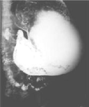
One hour after start of UGI
Clue: Nature oflesion in thisorgan
Gastric outlet obstruction
1.
2.
3.
4.
5.
Gastric outlet obstruction
1.Peptic ulcer disease
a. Acute or chronic
2.Carcinoma
a. Primary, Metastatic (pancreatic)
3.Gastric volvulus
4.Antral diaphragm/web
5.Pyloric stenosis in children
Clue: Nature oflesion in thisorgan
Gastric dilatation
1.
2.
3.
4.
5.
Gastric dilatation
1.Diabetes
2.Electrolyte/acid-base imbalance
3.Neuromuscular disorder
4.Abdominal surgery
5.Abdominal trauma
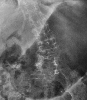
Clue: Nature oflesion in thisorgan
Widening of retrogastric space
1.
2.
3.
4.
Widening of retrogastric space
1.Pancreatic mass
2.Retroperitoneal mass
3.Exophytic posterior wall gastric tumor
4.Aortic aneurysm
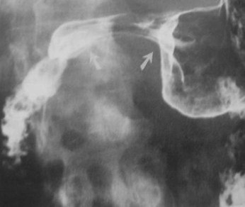
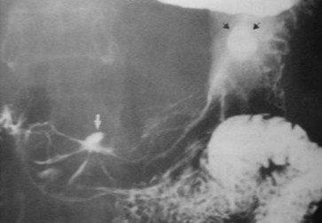
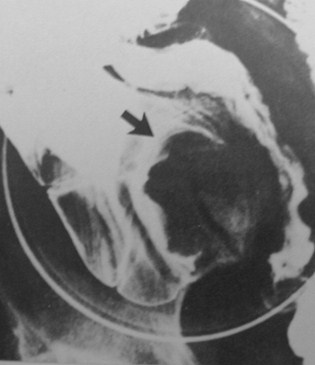
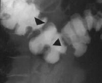
Clue: Type oflesion arrows arepointing to in thislocation
Antral Pad Sign
1.
2.
3.
Antral Pad Sign
1.Pancreatic cancer
2.Pancreatic pseudocyst
3.Gallbladder (normal or distended)
Clue: Extent oflesion
Diseases that cross the pylorus
1.
2.
3.
4.
5.
6.
1.Lymphoma
2.Carcinoma
3.Crohn’s Disease
4.Peptic ulcer disease
5.TB
6.Eosinophilic Gastroenteritis
Diseases that cross the pylorus
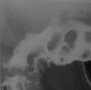
Clue: type oflesion in thisorgan
Multiple Duodenal Filling Defects
1.
2.
3.
Multiple Duodenal Filling Defects
1.Brunner’s gland hyperplasia
2.Polyps
1.Adenomatous, hyperplastic
3.Lymphoma
Clue: type oflesion in thisorgan
Thickened Duodenal Folds
1.
2.
3.
4.
5.
6.
Thickened Duodenal Folds
1.Duodenitis
2.Chronic Renal Failure
3.Pancreatitis
4.Zollinger-Ellison Syndrome
5.Varices
6.Lymphoma
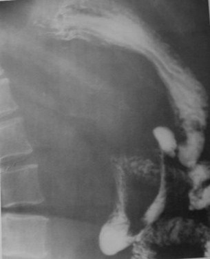
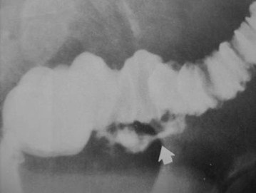
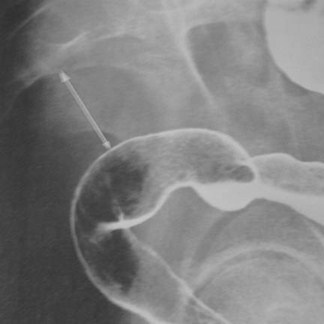
Clue: type oflesion
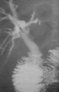
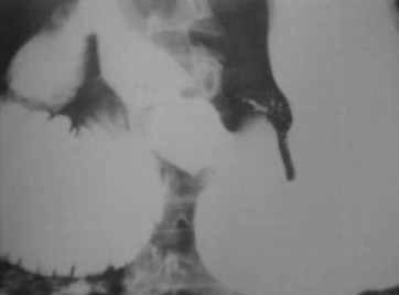
Duodenal-Biliary fistula
1.
2.
3.
4.
Duodenal-Biliary fistula
1.Prior sphincterotomy
2.Cholecystitis
3.Gallbladder carcinoma
4.Duodenal carcinoma
Clue: type oflesion in thisorgan
Duodenal Obstruction
1.
2.
3.
4.
5.
6.
Duodenal Obstruction
1.Congenital duodenal atresia/stenosis
2.Annular pancreas
3.Midgut volvulus
4.Carcinoma
5.SMA syndrome
6.Intramural hematoma
Clue: type oflesion in thisorgan


Widening of duodenal sweep
1.
2.
3.
4.
Widening of duodenal sweep
1.Pancreatic Disease
a. Pancreatitis, pseudocyst, tumor
2.Lymphadenopathy
3.Aortic Aneurysm
4.Choledochal cyst
Clue: type oflesion in thisorgan
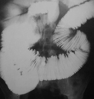
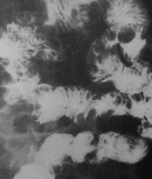
Dilated small bowelNo fold thickening
1.
2.
3.
Dilated small bowelNo fold thickening
1.Scleroderma
2.Sprue
3.Small Bowel Obstruction
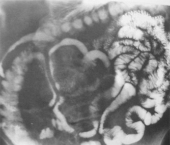
Clue: type oflesion in thisorgan
Multiple small bowel masses
1.
2.
3.
Multiple small bowel masses
1.Polyposis (Peutz-Jeghers)
2.Lymphoma
3.Metastases
Clue: type oflesion in thisorgan
Separation of small bowel loops
1.
2.
3.
4.
5.
6.
Separation of small bowel loops
1.Thickening of bowel wall
2.Carcinoid
3.Crohn’s Disease
4.Ascites
5.Peritoneal metastases
6.Retractile mesenteritis
Clue: type oflesion in thisorgan
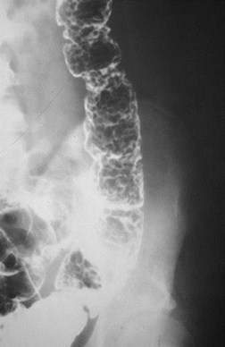
Multiple Colonic Filling Defects
1.
2.
3.
4.
Multiple Colonic Filling Defects
1.Polyps (polyposis)
2.Pseudopolyps (UC)
3.Lymphoma
4.Cystic Pneumatosis
Clue: type oflesion in thisorgan
Smooth Colonic Stricture
1.
2.
3.
4.
Smooth Colonic Stricture
1.Ulcerative Colitis
2.Crohn’s Disease
3.Radiation
4.Ischemia
Clue: type oflesion in thisorgan
Ileocecal disease
1.
2.
3.
4.
Ileocecal Disease
1.Crohn’s Disease
2.Lymphoma
3.TB
4.Cancer(colon adenocarcinoma, carcinoid)
Clue: type oflesion in thisorgan
Coned Cecum
1.
2.
3.
Coned Cecum
1.Amebiasis
2.Crohn’s Disease
3.TB
Clue: type oflesion in thisorgan
Cecal Filling defect
1.
2.
3.
4.
5.
Cecal Filling defect
1.Appendicitis/Mucocele
2.Crohn’s Disease
3.Cecal neoplasm
4.Ileocolic intussusception
5.Lymphoma
Clue: type oflesion in thisorgan
Double tracking of Colon
1.
2.
3.
Double tracking of Colon
1.Crohn’s Disease
2.Diverticulitis
3.Adenocarcinoma
Clue: type oflesion in thisorgan


Colon Thumbprinting
1.
2.
3.
4.
Colon Thumbprinting
1.Ulcerative Colitis
2.Ischemia
3.Hemorrhage
4.Lymphoma
Clue: type oflesion in thisorgan
Smooth Colon
1.
2.
3.
4.
Smooth Colon
1.Ulcerative colitis
2.Cathartic Colon
3.Post ischemic
4.Post radiation
Clue: type oflesion in thisorgan

Widened Presacral space
1.
2.
3.
4.
5.
Widened Presacral space
1.Ulcerative colitis
2.Pelvic lipomatosis
3.Crohn’s Disease
4.Rectal tumor
5.Sacral tumor
Clue: type oflesion in thisorgan
Rectal Narrowing
1.
2.
3.
4.
5.
Rectal Narrowing
1.Ulcerative Colitis
2.Pelvic Lipomatosis
3.Lymphogranuloma Venereum (LGV)
4.Solitary Rectal Ulcer Syndrome
5.Radiation
The End