
Thyroid Masses
© William Herring, MD, FACR
Anterior Mediastinal Masses
Thyroid
Thymoma
Teratoma
Lymphoma
Substernal Thyroid
Substernal ThyroidLocation
Most (80%) arise from lower pole or isthmusof thyroid and extend into anteriormediastinum
Some (20%) arise from posterior aspect ofthyroid and extend into posteriormediastinum
Almost always on right
Substernal ThyroidClinical
Most patients are asymptomatic
Thyrotoxicosis and carcinoma rare
Substernal Thyroid Pathology
Usually nodular, colloid goiters
Typically well-encapsulated
May show degeneration (calcification)
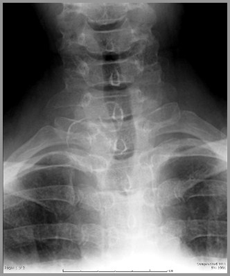
Substernal ThyroidImaging-1
Sharply defined, smooth or lobulated softtissue mass
Characteristically displaces trachea
Usually do not project below arch of aorta
Differentiates them from thymomas andteratomas
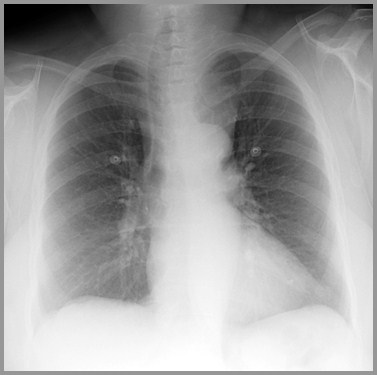
Substernal thyroid displaces thetrachea to the right
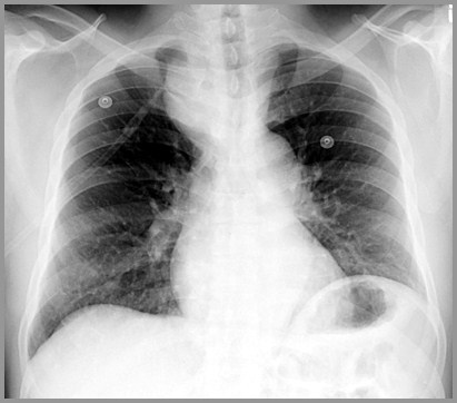
Substernal thyroid displaces thetrachea to the left
Substernal ThyroidImaging-2
When they occur posteriorly, theycharacteristically interpose betweentrachea in front and esophagus in back
Curvilinear calcifications highlysuggestive of a degenerated thyroidadenoma

Two thyroid adenomas withcurvilinear calcification
Substernal ThyroidCT Appearance
Usually contrast enhance and manytimes contain calcification
Contrast enhancement is prolonged
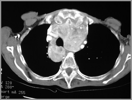
Substernal thyroid enhances with contrastand displaces trachea to the right
Substernal ThyroidDiagnosis
Radioisotope scan is diagnostic
The End