
PosteriorMediastinal Masses
PosteriorMediastinal Masses
© William Herring, MD, FACR
Neurogenic Tumors
Peripheral nerve origin
Neurofibromas
Neurilemomas (Schwannomas)
Neurogenic Tumors
Sympathetic nerve origin
GanglioneuromasUsually benign
NeuroblastomasUsually malignant
SympathicoblastomasUsually malignant
Paraganglionic cells
Pheochromocytoma
Chemodactomas (paragangliomas)—benign ormalignant
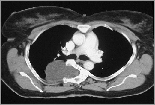
Posterior MediastinumAnatomy
Bordered anteriorly by pericardium
Posteriorly by anterior border of spine
Paravertebral gutters technically excluded fromposterior mediastinum
For practical purposes considered part of it
Posterior MediastinumContents
Descending aorta
Esophagus
Thoracic duct
Vagus nerves
Nodes
Posterior Mediastinal MassesBenign vs. Malignant
About 30% malignant
Nerve sheath tumors most common andusually benign
Neoplasms which arise from nerveelements other than sheath usuallymalignant
Posterior Mediastinal MassesNeurofibromas
In adults, neurofibroma and neurilemomas(Schwannomas) most common
Neurofibroma contains Schwann cells plusnerve cells
Occur 3rd or 4th decade
Schwannoma derived from sheath of Schwannwithout nerve cell
Posterior Mediastinal MassesGanglioneuroma
In children, ganglioneuroma andneuroblastoma are most common
Ganglioneuromabenign tumor
Neuroblastomamalignant tumor
Posterior Mediastinal MassesNeuroblastoma
Very malignant, undifferentiated roundcell lesion
From sympathetic ganglion
Usually <10 years old
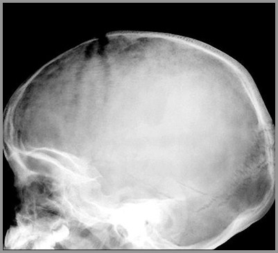
Produces “iron-filings” appearance tosutures in skull infiltrated with tumor
“Iron Filings” appearance of suture from metastases from neuroblastoma
“Iron Filings” appearance of suture from metastases from neuroblastoma
Posterior Mediastinal MassesCalcification
Calcification in posterior mediastinalmass points to neural tumor in childrather than met from somewhere else
Posterior Mediastinal MassesGeneral
Posterior mediastinal neurofibromasonly rarely associated withneurofibromatosis
Most have no symptoms
Posterior Mediastinal MassesImaging
Rib erosions
Both benign and malignant
Enlarged neural foramina (dumbell shapedlesion)
Scalloping of posterior vertebral bodies
Scoliosis
Pleural effusion
Both benign and malignant neural tumor
NeurofibromaMultiple subcutaneous neurofibromas (yellow arrows)
NeurofibromaMultiple subcutaneous neurofibromas (yellow arrows)
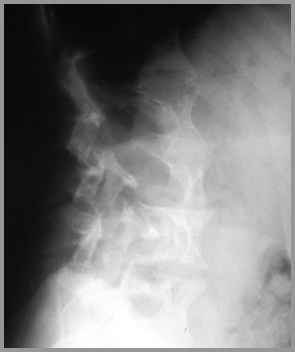
Posterior Scalloping of lumbarvertebral bodies in neurofibromatosis
Posterior Scalloping of lumbarvertebral bodies in neurofibromatosis
Lateral Meningocele in neurofibromatosis
Lateral Meningocele in neurofibromatosis
Posterior Mediastinal MassesParaspinal Abscess
Paraspinal abscess from TB – look fordestruction of two contiguous endplatesplus narrowing of intervening disc space
Posterior Mediastinal MassesOther Causes
Neurenteric cysts may have associatedhemivertebra
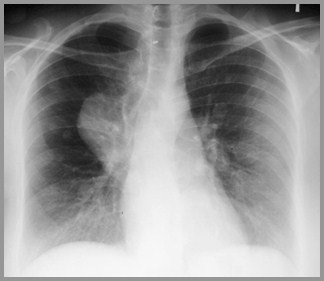
Extramedullary hematopoiesis should beassociated with splenomegaly andsometimes widening of ribs
Posterior mediastinal mass from extramedullary hematopoiesis
Posterior mediastinal mass from extramedullary hematopoiesis
The End
The End