|
|
Imaging Findings of Cirrhosis
Hepatic cirrhosis is the clinical and pathologic result of chronic liver injury, multifactorial in etiology, producing extensive fibrosis and nodular regeneration replacing the normal liver parenchyma. Cirrhosis is associated with a markedly increased risk of hepatocellular carcinoma (HCC).
CT
- Nodularity of the surface of the liver
- Hypertrophy of the caudate lobe as well as the lateral segments of the left lobe
- Atrophy of the posterior segments of the right lobe
- Fatty infiltration
- Heterogeneous appearance of the liver parenchyma pre- and post-contrast
- Regenerative nodules that may be isodense or hyperdense.
- Signs of portal hypertension which can include portal vein enlargement and portal venous thrombosis
- Intrahepatic vascular shunting manifest as early opacification of the intrahepatic veins during the early arterial phase-injection often associated with geographic, wedge-shaped perfusion abnormalities
- Splenomegaly
- Ascites
- Varices
Ultrasound
- Surface nodularity
- Diffuse coarse and heterogeneous echo pattern
- Fatty infiltration resulting in increased echogenicity which may be diffuse or focal
- Hypertrophy of the caudate lobe as well as the lateral segments of the left lobe
- Atrophy of the posterior segments of the right lobe
- Signs of portal hypertension including
- Enlarged portal vein
- Enlarged superior mesenteric vein and splenic vein
- Reversal of to-and-fro portal venous flow
- Portal vein thrombosis
- Transformation of the hepatic vein waveform to a portal vein waveform
- Portosystemic collaterals
- Splenomegaly
- Ascites
- Varices
MRI
Major role in identifying hepatocellular carcinoma (HCC)
- Nodularity of the surface of the liver
- Hypertrophy of the caudate lobe (more often in alcoholic cirrhosis) as well as the lateral segments of the left lobe
- Atrophy of the posterior segments of the right lobe
- Fatty infiltration
- Heterogeneous appearance of the liver parenchyma pre- and post-contrast
- Expanded gallbladder fossa sign
- Enlargement of the pericholecystic space
- Regenerative nodules (or cirrhotic nodules)
- T1 variable, usually isointense. Since supply is from portal vein, no early enhancement or washout
- T2 usually isointense
- Dysplastic nodules may have variable appearance
- Low-grade nodules resemble regenerative nodules
- High-grade nodules resemble HCCs
- Hepatocellular carcinoma (HCC)
- T1 usually hyperintense, with early arterial enhancement and washout.
- T2 is typically hyperintense
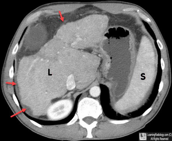
Cirrhosis, CT. The liver (L) is shrunken in size and has a nodular margin (red arrows). The spleen (S) was enlarged.
MedScape. Cirrhosis Imaging. CR Taylor.
MDCT Imaging Findings of Liver Cirrhosis: Spectrum of Hepatic and Extrahepatic Abdominal Complications. GP Sangster, CH. Previgliano, M Nader, E Chwoschtschinsky, and MG Heldmann. HPB Surgery. Volume 2013 (2013).
|
|
|
