|
|
Reactive Airways Disease
General Considerations
- General term for a disease usually in the pediatric population featuring wheezing, shortness of breath and coughing
- Initial episodes are frequently referred to as bronchiolitis
- Unlike asthma, which is chronic, reactive airways disease is usually transient although it can progress over time to asthma
- May be triggered by
- Viral URIs, especially from respiratory syncytial virus (RSV)
- Pollen and mold
- Cigarette smoke
- Extreme cold
- Most (60%) of children who have wheezing before age 3 will outgrow it by age 6
- Use of the term is controversial, some believing it is too general
Clinical Findings
- Increased respiratory rate
- Retractions
- Cough
- Fever
- Rhinorrhea
Imaging Findings
- Peribronchial thickening
- Primarily lobar or segmental bronchi
- While adults may have bronchi on end visible in the hila, children usually do not
- Peribronchial thickening also produces tram-track like linear densities in the lung from bronchi visualize in profile
- Hyperinflation
- Atelectasis from mucus plugging
Differential Diagnosis
- It is usually impossible to distinguish between viral bronchiolitis and asthma in a young child and the two may coexist
- Reactive airways dysfunction syndrome (RADS) and irritant-induced asthma (IrIA)
- Closely related forms of asthma that result from the nonimmunologicprovocation of prolonged bronchial hyperresponsiveness and airflow obstruction by inhaled irritants
- Anaphylactic reaction
- Foreign body aspiration
Treatment
- Bronchodilator
- Steroids
- Oxygen


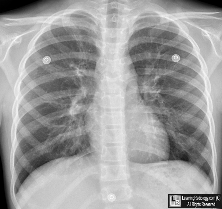
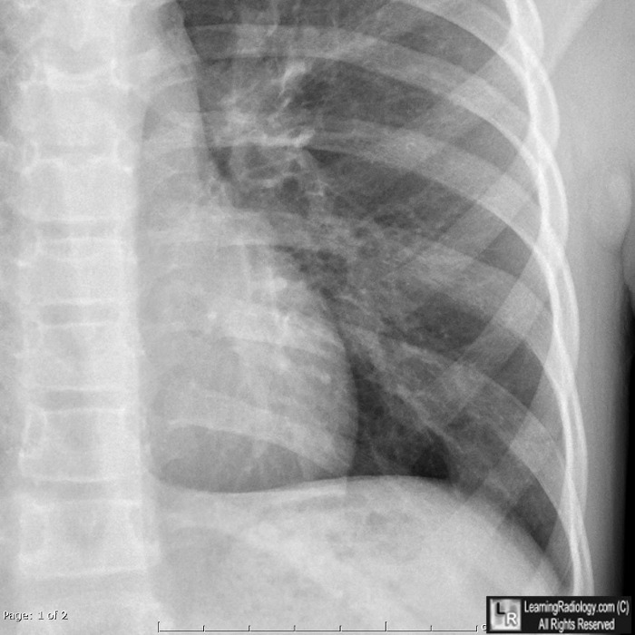
Reactive Airways Disease. (Top) Peribronchial thickening (white circles) seen en face shows small donut-like rings in periphery of lungs, not normally seen. Contained in yellow circle are thickened bronchial walls seen in profile with a "tram-track appearance. (Bottom) Close-up of left lower lung in same patient shows more donut shaped thickened bronchial walls. (yellow arrows)
For these same photos without the arrows, click here and here
For more information, click on the link if you see this icon 
Imaging in Pediatric Pulmonology, edited by RH Cleveland
|
|
|
