|
|
Heterotopic Ossification
General considerations
- Defined as the abnormal formation of true bone within extra-skeletal soft tissues
- More common in males, especially following spinal cord injury, it is rare in young children
- Formerly called myositis ossificans
- That term has fallen out of favor because the condition is not always inflammatory and ossification occurs in soft tissues other than muscle
- Strong association exists between HO and spinal cord or traumatic brain injury
- About 20-30% of patients with neurologic deficits will develop HO, possibly higher with spinal cord injuries
- It is also seen in burn patients, following surgery, and following blunt trauma such as horse riders may develop in the adductor muscles of the leg
- There is an increased risk for HO in patients with Diffuse Idiopathic Skeletal Hyperostosis (DISH) and Paget’s Disease
Pathophysiology
- Bone morphogenetic proteins may stimulate primitive stem cells in soft tissues to form osteoblasts under certain conditions
- Following trauma, cartilage begins developing in soft tissues by 2nd week, with trabeculated bone appearing by 2-5 weeks
Clinical findings
- May cause pain and a palpable mass
- Contributes to further restricting range of motion
- Can lead to breakdown of the skin in spinal cord patients
- Post-surgical HO most commonly occurs at hip following arthroplasty which is also the most common site of HO in patients with brain or spinal cord injury
- Shoulders and elbows follow in frequency in brain injury
- Knees are uncommonly affected in brain injury but frequently affected in spinal cord injury
Imaging findings
- Conventional radiography is the study of first choice
- Nuclear bone scan is most sensitive for early detection
- Bone can be detected on conventional radiographs as early as two weeks after injury, the ossification typically starting at the periphery as a cloud-like increase in density
- Biopsy of the lesion could lead to a false-positive diagnosis of osteosarcoma unless the clinical findings are taken into account and time is allowed for the lesion to mature
- CT may show a soft tissue mass early, followed by visualization of bone earlier than can be seen with conventional radiographs
- MRI is typically not used
- Ultrasound may show abnormalities in the muscle in advance of visible ossification
- The standard for early HO detection is the triple-phase bone scan using Tc 99M MDP
- Bone scans may be positive 2-6 weeks earlier than ossification is visible
- Early in the course, only the blood pool images may be positive whereas abnormal uptake during the soft tissue phase is diagnostic later in the course of the disease
Treatment
- Prophylactic treatment may include radiation therapy using an external beam, non-steroidal inflammatory agents and oral etidronate
- Surgical resection, when performed, is usually done only after the lesion has matured, the progress of which can be monitored by bone scan
- Recurrence is relatively common following resection

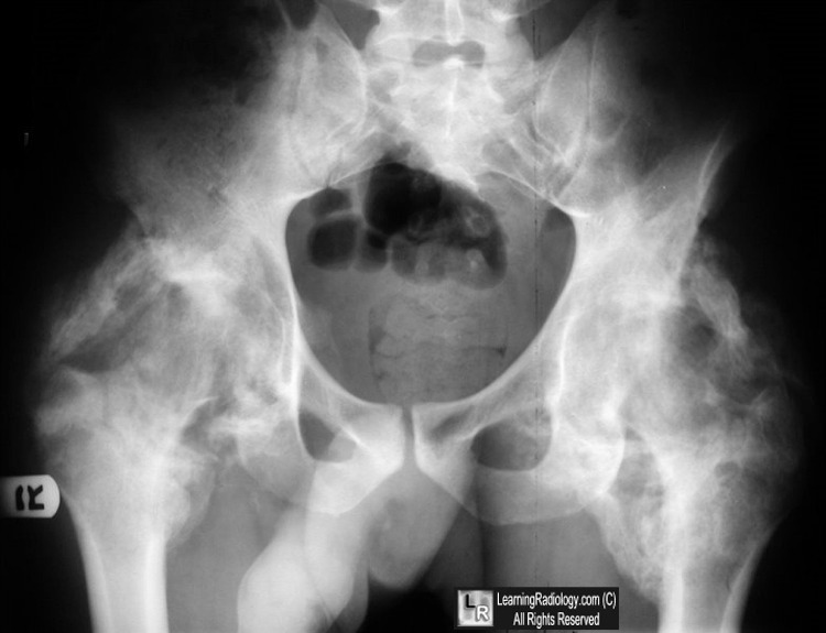
Heterotopic ossification. White arrows point to ossification (with trabeculae and cortex)
surrounding both hip joints in a young patient with a traumatic brain injury several months earlier
For this same photo without arrows, click here
For more information, click on the link if you see this icon 
|
|
|
