|
|
Medullary Nephrocalcinosis
General Considerations
- Increased calcium content of kidneys
- Usually applies to a generalized, rather than localized, increase in renal calcium content
- Macroscopic nephrocalcinosis is nephrocalcinosis visible without magnification discovered by radiography, ultrasonography, or at autopsy
- Macroscopic nephrocalcinosis can affect either cortex or medulla
- Medulla more common
- Diffuse calcification rarely observed in chronic glomerulonephritis or long-standing chronic renal disease
- Cortical nephrocalcinosis is rare and usually occurs secondary to diffuse cortical disease
- Calcification can be patchy or confluent
Clinical findings
- Calcium nodules commonly rupture through the papillary epithelium into the calyceal system to become urinary stones and, therefore, the presentation may be that of
- Renal colic
- Hematuria
- Urinary tract infection
- Passage of urinary stones
- Macroscopic nephrocalcinosis should not be considered synonymous with urinary stones because it signifies a metabolic derangement and has broader implications
- Episodes of urinary tract infections may occur
- Polyuria and polydipsia may be prominent
- Hypertension less common
- Microscopic pyuria invariably found
- Represents chronic inflammatory response to medullary calcification
- Distal tubular dysfunction is common with a mild salt-losing defect
Imaging Findings
- Medullary nephrocalcinosis
- Small nodules of calcification clustered in each pyramid
- Diagnosing underlying renal disease based on the appearance of calcification is difficult, except in the cases of:
- Papillary necrosis due to analgesic abuse because the entire papilla may be calcified, and
- Medullary sponge kidney where the sharp areas of calcification and uneven distribution may be seen
Causes
- Primary hyperparathyroidism is single most common cause of nephrocalcinosis in adults
- Nephrocalcinosis related more to the duration than the intensity of hypercalcemia
- Nephrocalcinosis occurs in 5% of the cases of hyperparathyroidism
- Distal Renal Tubular Acidosis is second most common cause of medullary nephrocalcinosis
- Both familial and secondary forms have high incidence
- Contributing mechanisms are hypercalcemia, acidosis, and reduced excretion of citrate in the presence of increased urinary pH
- Renal function is fairly well maintained
- Other causes of nephrocalcinosis are
- Hypervitaminosis D due to treatment of hypoparathyroidism or self-administration of vitamins
- Milk-alkali syndrome due to ingestion of milk or alkali for ulcer dyspepsia
- Sarcoidosis due to increased conversion of 25-hydroxycholecalciferol to 1,25-dihydroxycholecalciferol within the sarcoid granuloma
- In children with hypophosphatemic rickets, nephrocalcinosis increasingly being recognized as most common complication
- Idiopathic hypercalcuria, one of the common metabolic diseases, also is known cause of nephrocalcinosis
- Medullary sponge kidney is a common cause of medullary calcification in which calcium lies in ectatic collecting ducts rather than renal substance
- Calcium deposits are larger and more sharply defined than in metabolic disease
- They are uneven in distribution
- Associated hemihypertrophy of the body may exist
- Nephrocalcinosis associated with distal RTA and medullary sponge kidney usually is gross and renal function is relatively well preserved
- Renal papillary necrosis associated with phenacetin-induced analgesic nephropathy is identified as calcified papillae rather than speckled pattern.
- Rapidly progressive osteoporosis due to immobilization, menopause, senility, or steroids also may cause nephrocalcinosis
- Hyperoxaluria, primary (familial) or secondary to increased intake of oxalates, enhanced absorption due to intestinal disease, or ingestion of ethylene glycol or methoxyflurane can induce medullary calcification
Prognosis
- Depends mainly on the etiology of nephrocalcinosis
- Major long-term complication in patients with medullary nephrocalcinosis is renal failure
- Early treatment of reversible causes of renal failure, such as treatment of urinary infections, calculus obstruction, and hypertension, is essential
- Once renal failure is established, it must be treated accordingly
- Patients with idiopathic hypercalcuria and medullary sponge kidney have the least risk of renal failure and the best prognosis, whereas patients with primary type 1 hyperoxaluria have the worst prognosis


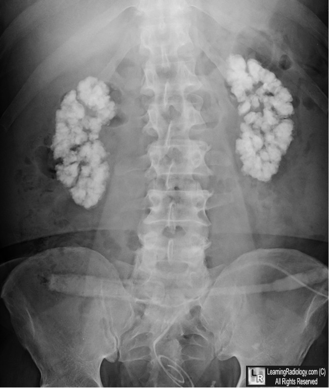
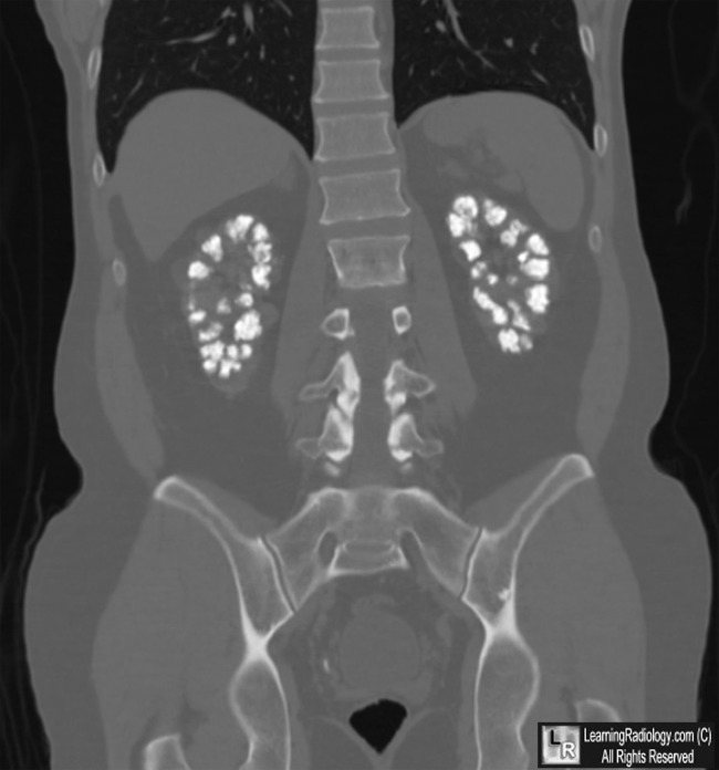
Medullary Nephrocalcinosis. Upper photo of conventional radiograph of abdomen and lower photo
of coronal CT scan of abdomen both show amorphous, coarse calcifications throughout both kidneys
(white arrows) which correspond the the shape and position of the renal pyramids. The patient had renal tubular acidosis.
For these same photos without arrows, click here and here
For more information, click on the link if you see this icon 
Mahendra Agraharkar, MD, FACP, President, Space City Associates of Nephrology; Medical Director, Acute Dialysis Unit and Chronic Home Dialysis Unit, Gambro Healthcare Reliant Dialysis Center
Coauthor(s): Rajiv Gupta, MD, Fellow, Department of Medicine, Division of Cardiology, John Sealy Hospital, University of Texas Medical Branch at Galveston
eMedicine Nephrocalcinosis
|
|
|
