|
|
Patent Ductus Arteriosus
PDA
General Considerations
- Persistent communication between the thoracic aorta and the pulmonary artery by the ductus arteriosus
- In fetal life, ductus carried blood from the pulmonary artery to the aorta, essentially bypassing the fetal lungs
- The ductus usually closes functionally within a few hours after birth
- If the ductus remains patent beyond 3 months, it is considered abnormal
- The effect of the left-to-right shunt will depend on the size of the shunt and the pulmonary vascular resistance
- If ductus persists, the shunt will be left-to-right from the aorta to the pulmonary artery
- May be an obligatory shunt in complex cardiac lesions
- Hypoplastic left heart syndrome
- D-Transposition
- Pulmonary atresia
Clinical Findings
- Although presentation can be at any age, PDA usually presents in childhood
- If shunt is large, may present with congestive heart failure
- Acyanotic, until or unless Eisenmenger’s physiology leads to reversal of the flow to right-to-left
- Increased pulmonary infections
- Inability or difficulty with feeding
- Weight loss (or no weight gain)
Imaging Findings
- The diagnosis is based on clinical findings, including EKG, imaging and echocardiographic findings, the latter being the primary means of imaging the lesion
- Chest radiographs yield non-specific findings such as CHF, a large main pulmonary artery and increased shunt vasculature
- The ductus may be long or short, relatively straight or tortuous
- Tends to be wider on the aortic side
- Forms acute angle with aorta in isolated PDA; more obtuse angle with associated congenital heart disease
- When closed, the ductus forms the ligamentum arteriosus, which may calcify in aorto-pulmonary window
- MRI findings
- Usually cardiac-gated T1 weighted (black blood) imaging
- Sagittal oblique plane through aortic arch shows ductus
Complications
- Aortic rupture
- Eisenmenger physiology
- Left heart failure
- Myocardial ischemia
- Necrotizing enterocolitis
- Pulmonary hypertension
Treatment
- If administered within two weeks of birth, intravenous indomethacin or IV ibuprofen are often effective in closing a PDA
- Catheter closure
- Surgical ligation
- It may be desirable to keep the ductus patent (as in cyanotic heart disease) in which case Prostaglandin E1 can be used
Prognosis
- Generally considered excellent in patients in whom the PDA is an isolated abnormality


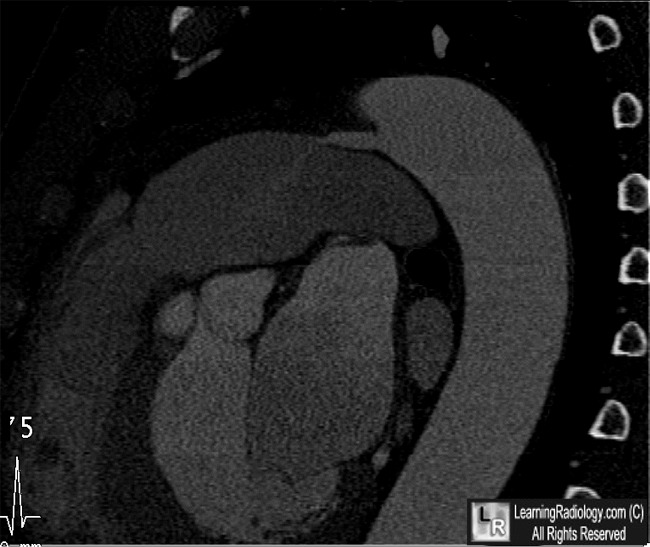
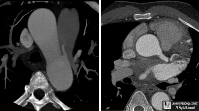
Patent Ductus Arteriosus (PDA). Upper: Sagittal reconstruction of contrast-enhanced cardiac CT shows a tubular communication (yellow arrow) between the Aorta (Ao) and the pulmonary artery (PA). A wisp of more slightly enhancing contrast is seen coming from the PDA into the PA (white arrow). Lower: Axial views again show the tubular PDA (yellow arrows) emanating from the aorta (AO).
For these same photos without the arrows, click here and here
For more information, click on the link if you see this icon 
|
|
|
