|
|
Acetabular Protrusio
Protrusio Acetabuli
General Considerations
- Intrapelvic displacement of the medial acetabular wall
- Most common cause is osteoarthritis
- Primary form – Otto pelvis
- Marked female to male predominance
- Usually occurs in young to middle-aged
- Bilateral in 1/3 to 2/3 of patients
- No underlying causative mechanism is demonstrated
- Secondary form
- Rheumatoid arthritis
- Paget disease
- Central fracture-dislocation
- Hip implants
- Marfan Syndrome
- Osteomalacia
Clinical Findings
- May be asymptomatic, or have
- Limitation of motion
- Joint stiffness
- Pain
Imaging Findings
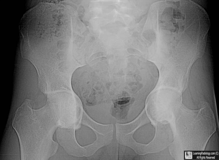
- Bilateral axial migration of the femoral heads with or without moderate degenerative changes
- Distance between the acetabulum and the ilioischial line should be >3 mm in males and >6 mm in females in protrusio
- Femoral head should not project medial to a line joining the inner border of the pelvis and the lateral margin of the obturator foramen
Complications
- Coxa vara and decreased femoral anteversion
Treatment
- Depends on age and degree of degenerative changes
- Medial wall bone grafts
- Joint replacement surgery may be necessary

Acetabular Protrusio. There is bilateral acetabular protrusio White arrows). The femoral head should not extend medial to a line
drawn from the lateral aspect of the pelvis and the lateral aspect of the obturator foramen (blue line). The distance between
the acetabulum and the ilioischial line (yellow arrow) should not be > 3mm in males and >6 mm in females.
For this same photo without the arrows, click here
For more information, click on the link if you see this icon 
Protrusio Acetabuli. CC Dunlop, CW Jones, and N Maffulli. Bulletin, Hospital for Joint Diseases Volume 62, Numbers 3 & 4 2005
|
|
|
