|
|
Epidermal Inclusion Cyst
Epidermoid Cyst of Bone
General Considerations
- Associated with trauma, especially penetrating trauma
- Usually in bones that are superficially located such as fingers, foot and calvarium
- Implantation of epithelium that form cysts leading to bone erosion
- Subungual crush-type injuries have been associated with inclusion cysts as has prior surgery
Clinical Findings
- May develop with in weeks or years of fingertip injury
- Pain at site of lesion
- Mass
Imaging Findings
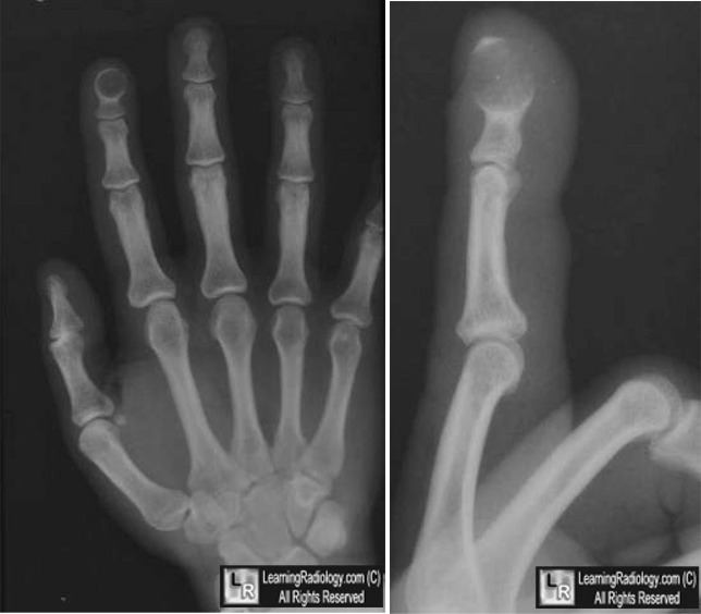
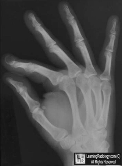
- Frequently seen in terminal phalanx
- Solitary, lytic lesion
- May be expansile
- May have a thin sclerotic border
Differential Diagnosis
- Enchondroma
- Metastases (rare)
- Glomus tumor
Treatment
Complications
- May recur if incompletely curetted
Prognosis


Epidermal Inclusion Cyst. White arrows pint to an expansile lytic lesion in the terminal phalanx
of the index finger in a characteristic location for an epidermal inclusion cyst of the hand.
For these same photos without the arrows, click here and here
For more information, click on the link if you see this icon 
|
|
|
