|
|
Paratracheal Air Cysts
General Considerations
- Benign incidental finding on CT (rarely seen on chest radiographs)
- 2.0% to 3.7% of the population
- Most are probably tracheal diverticulum
- On imaging, 35% have a narrow stalk to the trachea
- Histology shows ciliated columnar epithelium
Clinical Findings
- None
- Not associated with either trauma or emphysema
Imaging Findings
- Always located on the right posterior trachea at the level of the thoracic inlet (~T2)
- Unilocular or multilocular air-filled cystic structures
- 2-20 mm in diameter on axial plane
- Frequently associated with contour abnormality of trachea
- May or may not see connection to trachea
Differential Diagnosis
- Herniated lung
- Remains in continuity with the lung
- Emphysematous blebs
- Laryngocele
- Mounier-Kuhn Syndrome

- Also has tracheomegaly and bronchiectasis
- Pneumomediastinum
- Air usually encircles trachea
Treatment


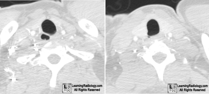
Paratracheal Air Cysts. Axial CT: There is a small pocket of air (blue and green arrows)
located to the right and slightly posterolateral to the trachea (T) at the levels of C7-T2.
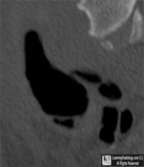
Sagittal reconstruction: Multi located collections of air (red arrow) communicate with the posterior aspect of the trachea (T) by a thin stalk (yellow arrow).
For these same photos without the arrows, click here and here
For more information, click on the link if you see this icon 
Buterbaugh JE, Erly WK. Paratracheal air cysts: a common finding on routine CT examinations of the cervical spine and neck that may mimic pneumomediastinum in patients with traumatic injuries. AJNR. 2008 Jun;29(6):1218-21
Goo JM, Im JG, Ahn JM, Moon WK, Chung JW, Park JH, Seo JB, Han MC. Right paratracheal air cysts in the thoracic inlet: clinical and radiologic significance. AJR. 1999 Jul;173(1):65-70
|
|
|
