|
|
Particle Disease
General Considerations
- AKA particle inclusion disease or giant cell granulomatous response or aggressive granulomatosis
- Occurs from inflammation and osteolysis secondary to the shedding of portions of a prosthesis, more often the polyethylene and/or methylmethacrylate cement in submicron size
- The granulomatous response elicited manifests as osteolysis
- Typically occurs 1-5 years after surgery, now most often in cementless prostheses
- The head may be made out of a cobalt-chromium alloy with a polyethylene cup
- Particles may migrate along the entire course of the prosthesis
Clinical Findings
- Asymptomatic until substantial bone loss
- Then, pain
- Limb shortening
- Limitation of motion
Imaging Findings
- Normal lucency is < 2mm at cement-bone interface
- Lucencies at metal-cement interface or metal-bone interface may be secondary to surgery and should remain unchanged over time
- They are usually 2 mm or less
- Lucencies greater than 2 mm can indicate loosening or infection or particle disease, or all three
- Particle disease usually produces multifocal lucencies which may not conform to the shape of the prosthesis
- There is usually no associated sclerotic reaction
- In the hip, the lesions occur mostly at the medial border of the tip of the femoral component
Differential Diagnosis
- Mechanical loosening
- Infection
Treatment
- Surgical revision is almost always necessary
Complications
- Dislocation
- Peri-prosthetic fracture


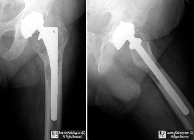
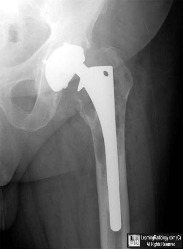
Particle Disease. The upper photos show a total left hip replacement 4 years after insertion demonstrating multiple lucencies (white arrows) surrounding the femoral portion of the prosthesis with endosteal scalloping. The lower photo shows progression of the disease with increased peri-prosthetic destruction 2 years later (yellow arrows).
For these same photos without the arrows, click here and here
For more information, click on the link if you see this icon 
Osteolysis and particle disease in hip replacement A review. William H Harris Acta Ortho~Scand 1994:65 113-123
|
|
|
