|
|
Milk of Calcium Bile
General Considerations
- Rare disorder in which gallbladder (GB) lumen is filled with a semi-solid, radio-opaque material
- Mostly calcium carbonate with calcium bilrubinate or calcium phosphate in a milky liquid
- Gallbladder stasis from longstanding cystic duct obstruction and cholelithiasis have been implicated in its formation
- It may be a long-term complication of total parenteral nutrition
- Predominately affects adults
- Milk of calcium may form in many other structures besides the gallbladder
Other Locations for Milk-of-Calcium |
Milk of calcium kidney cysts |
Calyceal diverticula |
Cysts in the breast |
Urethral diverticulum |
Bronchogenic cyst |
Mullerian duct cyst |
Clinical Findings
- May be asymptomatic
- The same as chronic cholecystitis
Imaging Findings
- Dense opacification of the gallbladder lumen seen on CT or conventional radiography
- Always associated with cholelithiasis or choledocholithiasis
- May form fluid-fluid level in conventional radiographs exposed with horizontal beam
- On ultrasound-intermediate density between sludge and gallstones
- Dense material settles In most dependent part of GB and may shadow
Differential Diagnosis
- Excretion of contrast into from a recent IV contrast injection
- Porcelain gallbladder
Treatment
- Surgical removal of GB is treatment of choice
- May pass spontaneously out of the GB
Complications
- Acute cholecystitis
- Pancreatitis
- Obstructive jaundice


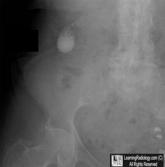
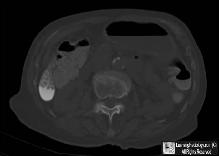
Milk of Calcium Bile. Conventional radiograph (above) shows milky substance in gallbladder (white arrow)
with multiple gallstones floating on top (black arrow). The CT scan (below) demonstrates
the same milk of calcium in the dependent part of the gallbladder (white arrow)
while the stones occupy the upper part (yellow arrow).
For these same photos without the arrows, click here and here
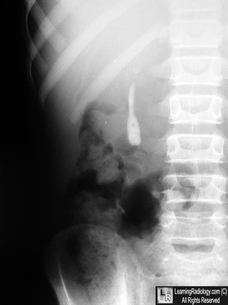
Milk of Calcium Bile. Conventional supine radiograph shows a dense, white, homogeneous structure in the shape of a shrunken gallbladder in the right upper quadrant.
For more information, click on the link if you see this icon 
|
|
|
